
Main menu

  • Online first
    • Online first
  • Current issue
    • Current issue
  • Archive
    • Archive
  • Submit a paper
    • Online submission site
    • Instructions for authors
  • About the journal
    • About the journal
    • Editorial board
    • Instructions for authors
    • FAQs
    • Chinese Stroke Association
  • Help
    • Contact us
    • Feedback form
    • Reprints
    • Permissions
    • Advertising
  • BMJ Journals

User menu

  • Login

Search

  • Advanced search
  • BMJ Journals
  • Login
  • Facebook
  • Twitter
Stroke and Vascular Neurology

Advanced Search

  • Online first
    • Online first
  • Current issue
    • Current issue
  • Archive
    • Archive
  • Submit a paper
    • Online submission site
    • Instructions for authors
  • About the journal
    • About the journal
    • Editorial board
    • Instructions for authors
    • FAQs
    • Chinese Stroke Association
  • Help
    • Contact us
    • Feedback form
    • Reprints
    • Permissions
    • Advertising
Open Access

New rat model of spinal cord infarction with long-lasting functional disabilities generated by intraspinal injection of endothelin-1

Masayuki Otani, Yoshihiro Kushida, Yasumasa Kuroda, Shohei Wakao, Yo Oguma, Keisuke Sasaki, Shintaro Katahira, Ryohei Terai, Rie Ryoke, Hiroi Nonaka, Ryuta Kawashima, Yoshikatsu Saiki, Mari Dezawa
DOI: 10.1136/svn-2023-002962 Published 21 June 2024
Masayuki Otani
1Division of Cardiovascular Surgery, Tohoku University Graduate School of Medicine, Sendai, Miyagi, Japan
2Department of Stem Cell Biology and Histology, Tohoku University Graduate School of Medicine, Sendai, Miyagi, Japan
  • Find this author on Google Scholar
  • Find this author on PubMed
  • Search for this author on this site
  • ORCID record for Masayuki Otani
Yoshihiro Kushida
2Department of Stem Cell Biology and Histology, Tohoku University Graduate School of Medicine, Sendai, Miyagi, Japan
  • Find this author on Google Scholar
  • Find this author on PubMed
  • Search for this author on this site
Yasumasa Kuroda
2Department of Stem Cell Biology and Histology, Tohoku University Graduate School of Medicine, Sendai, Miyagi, Japan
  • Find this author on Google Scholar
  • Find this author on PubMed
  • Search for this author on this site
Shohei Wakao
2Department of Stem Cell Biology and Histology, Tohoku University Graduate School of Medicine, Sendai, Miyagi, Japan
  • Find this author on Google Scholar
  • Find this author on PubMed
  • Search for this author on this site
Yo Oguma
2Department of Stem Cell Biology and Histology, Tohoku University Graduate School of Medicine, Sendai, Miyagi, Japan
  • Find this author on Google Scholar
  • Find this author on PubMed
  • Search for this author on this site
Keisuke Sasaki
3Department of Neurosurgery, Tohoku University Graduate School of Medicine, Sendai, Miyagi, Japan
  • Find this author on Google Scholar
  • Find this author on PubMed
  • Search for this author on this site
Shintaro Katahira
1Division of Cardiovascular Surgery, Tohoku University Graduate School of Medicine, Sendai, Miyagi, Japan
  • Find this author on Google Scholar
  • Find this author on PubMed
  • Search for this author on this site
Ryohei Terai
2Department of Stem Cell Biology and Histology, Tohoku University Graduate School of Medicine, Sendai, Miyagi, Japan
  • Find this author on Google Scholar
  • Find this author on PubMed
  • Search for this author on this site
Rie Ryoke
4Institute of Development, Aging, and Cancer, Tohoku University, Sendai, Miyagi, Japan
  • Find this author on Google Scholar
  • Find this author on PubMed
  • Search for this author on this site
Hiroi Nonaka
4Institute of Development, Aging, and Cancer, Tohoku University, Sendai, Miyagi, Japan
  • Find this author on Google Scholar
  • Find this author on PubMed
  • Search for this author on this site
Ryuta Kawashima
4Institute of Development, Aging, and Cancer, Tohoku University, Sendai, Miyagi, Japan
  • Find this author on Google Scholar
  • Find this author on PubMed
  • Search for this author on this site
Yoshikatsu Saiki
1Division of Cardiovascular Surgery, Tohoku University Graduate School of Medicine, Sendai, Miyagi, Japan
  • Find this author on Google Scholar
  • Find this author on PubMed
  • Search for this author on this site
Mari Dezawa
2Department of Stem Cell Biology and Histology, Tohoku University Graduate School of Medicine, Sendai, Miyagi, Japan
  • Find this author on Google Scholar
  • Find this author on PubMed
  • Search for this author on this site
  • Article
  • Figures & Data
  • eLetters
  • Info & Metrics
  • PDF
Loading

Article Figures & Data

Figures

  • Supplementary Materials
  • Additional Files
  • Figure 1
    • Download figure
    • Open in new tab
    • Download powerpoint
    Figure 1

    (A, B) The length of the silica tubing was adjusted to 1.5 mm using a digital calliper. (C) Schematic diagram of the intraoperative view. (D) Intraoperative picture under the microscope (see online supplemental movie 1). (E) Schematic diagram of the Evans blue injection centred on the anterior horn of the spinal cord (SC). (F) Representative example of Evans blue administration (0.7 µL) at angle X. (G) Overall image of the ventral side of the SC injected with 0.7 µL of Evans blue at each angle. (H) Diffusion of the dye in the SC, with 1.3 and 0.7 µL Evans blue. (I) Diffusion of the dye in the ventral SC was observed for two different volumes: 1.3 µL and 0.7 µL.
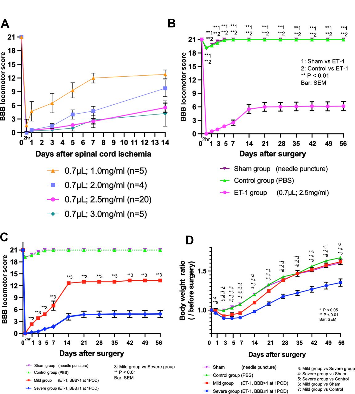
  • Figure 2
    • Download figure
    • Open in new tab
    • Download powerpoint
    Figure 2

    Changes in the BBB locomotor score and body weight ratio. (A) The volume of ET-1 was fixed at 0.7 µL/side, and the concentration was varied. Each rat was observed for 14 days, and mortality rates are shown below: all five rats/group administered 1.0 mg/mL and 3.0 mg/mL ET-1, respectively, survived for 14 days; 1 of the 5 rats administered 2.0 mg/mL ET-1 died on day 4, and the other rats survived for 14 days. Data for the rat that died during the observation period were excluded from the graph. (B) ET-1 (0.7 µL/side, 2.5 mg/mL) was injected. (C) The BBB score on postoperative day 1 predicted the BBB score in the chronic phase. At 2 hours postoperatively, both the severe and mild groups had a BBB score of 0. Already on day 1 after the injection, however, the BBB score differed significantly, with the severe group having a mean BBB score of 0.26±0.10 and the Mild group having a BBB score of 2.33±0.36 (p<0.01). (D) The body weight changes in each group up to day 56. The average preoperative weight was defined as 1.0. BBB, Basso, Beattie and Bresnahan; PBS, phosphate-buffered saline.

  • Figure 3
    • Download figure
    • Open in new tab
    • Download powerpoint
    Figure 3

    (A) TTC staining. Left: intact spinal cord (SC) at the Th13 level. Right: punctured cross-section on day 7n in the ET-1 group. (B) Vasoconstriction process after ET-1 injection. Blood flow at the level of Th13 was 61.6±7.4 mL/min/100 g before ET-1 administration, 15.9±4.0 mL/min/100 g immediately after ET-1 administration, 15.3±4.3 mL/min/100 g at 15 min, 31.9±1.4 mL/min/100 g at 30 min and 55.3±9.0 mL/min/100 g at 60 min. The blood flow was significantly decreased immediately, 15 min and 30 min after ET-1 administration compared with that before ET-1 administration (before vs just after; p<0.001, before vs 15 min; p<0.001, before vs 30 min; p<0.05). After 60 min, blood flow improved to the same level as before ET-1 administration (before vs 60 min; not significant). (C) Immunostaining with neurological markers, NeuN, Iba-1, GFAP and GST-pi. (D) Number of NeuN positive cells 3 days after ET-1 injection compared with intact SC. TTC, triphenyl tetrazolium chloride.

  • Figure 4
    • Download figure
    • Open in new tab
    • Download powerpoint
    Figure 4

    (A) TUNEL-positive cells per unit area (mm2) 3 days after ET-1 injection. (B) Double staining with TUNEL and NeuN. (C) Immunostaining for RECA-1. (D) RECA-1 positive vessels per unit area (mm2). The number of RECA-1 positive vessels (/mm2) in the anterior horn was 428.9±26.9 and 457.8±4.6 (p=0.38) and the number in the posterior horn was 505.6±17.6 and 503.3±23.1 (p=0.93) in the ET-1- (day 3) and intact groups, respectively. In the white matter, the number of RECA-1-positive vessels (/mm2) was 192.2±9.2 and 172.2±8.2 (p=0.11) on the ventral side and 188.9±10.0 and 163.3±6.6 (p=0.04) on the dorsal side in the ET-1- (day 3) and intact groups, respectively.

Supplementary Materials

  • Figures
  • Additional Files
  • Supplementary data

    [svn-2023-002962supp001.pdf]

  • Supplementary video

    [svn-2023-002962supp008.mp4]

  • Supplementary data

    [svn-2023-002962supp007.pdf]

  • Supplementary data

    [svn-2023-002962supp002.pdf]

  • Supplementary data

    [svn-2023-002962supp005.pdf]

  • Supplementary data

    [svn-2023-002962supp006.pdf]

  • Supplementary data

    [svn-2023-002962supp003.pdf]

  • Supplementary data

    [svn-2023-002962supp004.pdf]

Additional Files

  • Figures
  • Supplementary Materials
  • Supplementary Data

    This web only file has been produced by the BMJ Publishing Group from an electronic file supplied by the author(s) and has not been edited for content.

    • Data supplement 1
    • Data supplement 2
    • Data supplement 3
    • Data supplement 4
    • Data supplement 5
    • Data supplement 6
    • Data supplement 7
    • Data supplement 8
PreviousNext
Back to top
Vol 10 Issue 1 Table of Contents
Stroke and Vascular Neurology: 10 (1)
  • Table of Contents
  • Table of Contents (PDF)
  • About the Cover
  • Index by author
  • Ed Board (PDF)
Email

Thank you for your interest in spreading the word on Stroke and Vascular Neurology.

NOTE: We only request your email address so that the person you are recommending the page to knows that you wanted them to see it, and that it is not junk mail. We do not capture any email address.

Enter multiple addresses on separate lines or separate them with commas.
New rat model of spinal cord infarction with long-lasting functional disabilities generated by intraspinal injection of endothelin-1
(Your Name) has sent you a message from Stroke and Vascular Neurology
(Your Name) thought you would like to see the Stroke and Vascular Neurology web site.
CAPTCHA
This question is for testing whether or not you are a human visitor and to prevent automated spam submissions.
Print
Alerts
Sign In to Email Alerts with your Email Address
Citation Tools
New rat model of spinal cord infarction with long-lasting functional disabilities generated by intraspinal injection of endothelin-1
Masayuki Otani, Yoshihiro Kushida, Yasumasa Kuroda, Shohei Wakao, Yo Oguma, Keisuke Sasaki, Shintaro Katahira, Ryohei Terai, Rie Ryoke, Hiroi Nonaka, Ryuta Kawashima, Yoshikatsu Saiki, Mari Dezawa
Stroke and Vascular Neurology Jun 2024, svn-2023-002962; DOI: 10.1136/svn-2023-002962

Citation Manager Formats

  • BibTeX
  • Bookends
  • EasyBib
  • EndNote (tagged)
  • EndNote 8 (xml)
  • Medlars
  • Mendeley
  • Papers
  • RefWorks Tagged
  • Ref Manager
  • RIS
  • Zotero
Cite This
  • APA
  • Chicago
  • Endnote
  • MLA
Loading
New rat model of spinal cord infarction with long-lasting functional disabilities generated by intraspinal injection of endothelin-1
Masayuki Otani, Yoshihiro Kushida, Yasumasa Kuroda, Shohei Wakao, Yo Oguma, Keisuke Sasaki, Shintaro Katahira, Ryohei Terai, Rie Ryoke, Hiroi Nonaka, Ryuta Kawashima, Yoshikatsu Saiki, Mari Dezawa
Stroke and Vascular Neurology Jun 2024, svn-2023-002962; DOI: 10.1136/svn-2023-002962
Download PDF

Share
New rat model of spinal cord infarction with long-lasting functional disabilities generated by intraspinal injection of endothelin-1
Masayuki Otani, Yoshihiro Kushida, Yasumasa Kuroda, Shohei Wakao, Yo Oguma, Keisuke Sasaki, Shintaro Katahira, Ryohei Terai, Rie Ryoke, Hiroi Nonaka, Ryuta Kawashima, Yoshikatsu Saiki, Mari Dezawa
Stroke and Vascular Neurology Jun 2024, svn-2023-002962; DOI: 10.1136/svn-2023-002962
Reddit logo Twitter logo Facebook logo Mendeley logo
Respond to this article
  • Tweet Widget
  • Facebook Like
  • Google Plus One
  • Article
    • Abstract
    • Introduction
    • Materials and methods
    • Results
    • Discussion
    • Data availability statement
    • Ethics statements
    • Footnotes
    • References
  • Figures & Data
  • eLetters
  • Info & Metrics
  • PDF

Related Articles

Cited By...

More in this TOC Section

  • Stepwise improvement in intracerebral haematoma expansion prediction with advanced imaging: a comprehensive comparison of existing scores
  • Learning curve and embolisation strategy in single-stage surgery combined embolisation and microsurgery for brain arteriovenous malformations: results from a nationwide multicentre prospective registry study
  • Thrombus iodine-based perviousness is associated with recanalisation and functional outcomes in endovascular thrombectomy
Show more Original research

Similar Articles


CONTENT

  • Latest content
  • Current issue
  • Archive
  • eLetters
  • Sign up for email alerts
  • RSS

JOURNAL

  • About the journal
  • Editorial board
  • Recommend to librarian
  • Chinese Stroke Association

AUTHORS

  • Instructions for authors
  • Submit a paper
  • Track your article
  • Open Access at BMJ

HELP

  • Contact us
  • Reprints
  • Permissions
  • Advertising
  • Feedback form

© 2025 Chinese Stroke Association