

Article Figures & Data
Figures
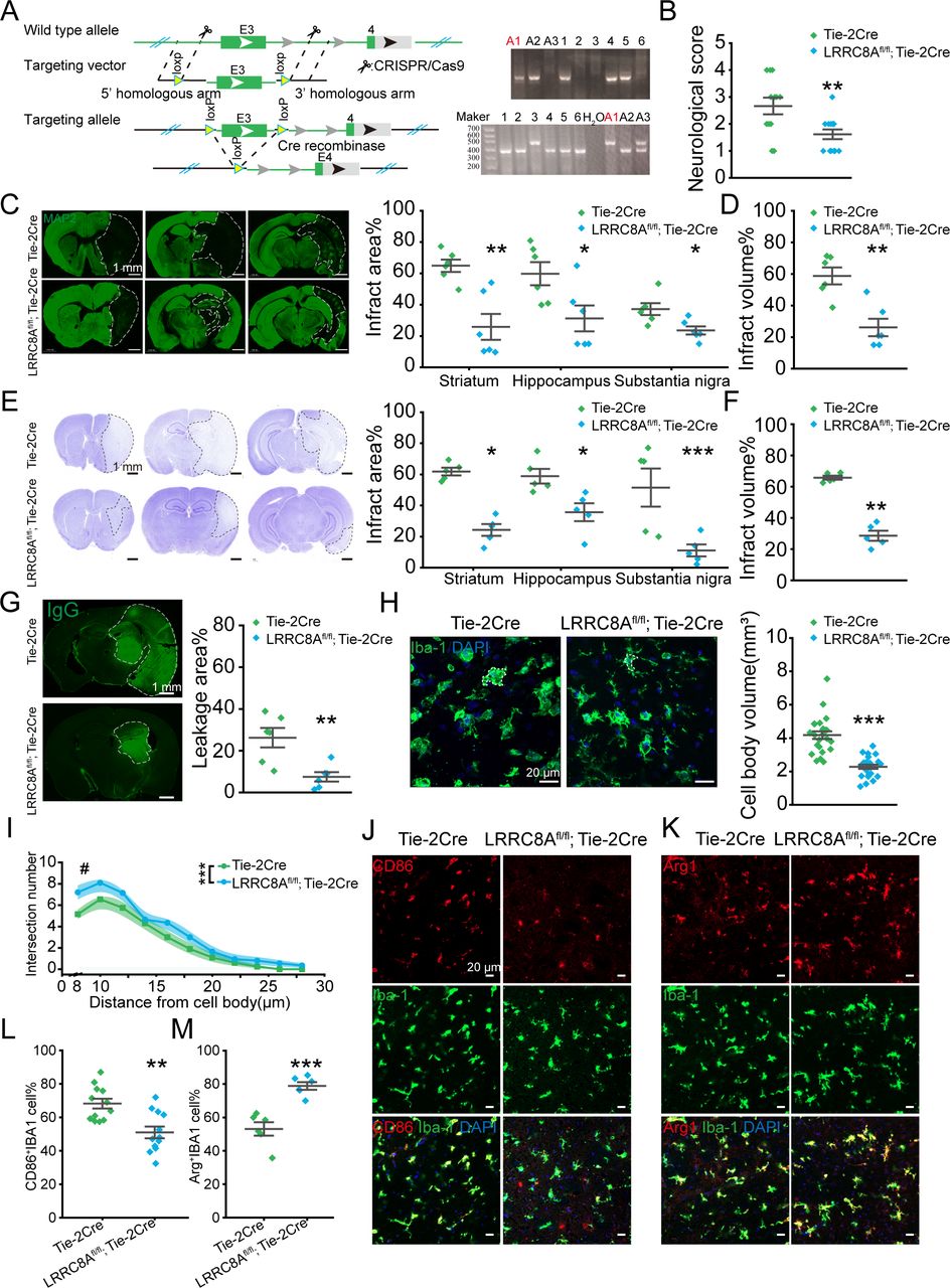
- Figure 1
Conditional knockout (cKO) of LRRC8A in endothelial cells could reduce brain injury post-ischaemic stroke. (A) Illustration of LRRC8A knockout with the CRISPR/Cas9 strategy. The right panel was the DNA genotype identification and the mice marked with ‘A1’ was the LRRC8Afl/fl; Tie-2Cre one. The genotype of LRRC8Afl/fl; Tie-2Cre was designated as the LRRC8A cKO mice and Tie-2Cre was the little-mate negative control. (B) The neurological scores 24 hours after ischaemia-reperfusion. N=13 mice in Tie-2Cre group, n=12 mice in LRRC8Afl/fl; Tie-2Cre group, **p<0.01, two-sample t-test. (C) Left panel: representative images of MAP2 immunostaining 24 hours after MCAO surgery. The white dotted lines indicated the borders of the infarct zones. The position of the brain slices from left to right was Bregma 0.86 mm, −1.34 mm and −2.80 mm. Right panel: quantification of the infarct areas across coronal sections. N=6 mice in Tie-2Cre group, n=6 mice in LRRC8Afl/fl; Tie-2Cre group, *p<0.05, **p<0.01, two-way ANOVA. (D) Quantification of total infarct volume. N=6 mice in the Tie-2Cre group, n=6 mice in the LRRC8Afl/fl; Tie-2Cre group, **p<0.01, two-sample t-test. (E) Left panel: representative images of Nissl staining 24 hours after MCAO. The white dotted lines indicated the borders of the infarct zones. The position of the brain slices from left to right was Bregma 0.86 mm, −1.34 mm and −2.80 mm. Right panel: quantification of the infarct areas across coronal sections. N=5 mice in Tie-2Cre group, n=5 mice in LRRC8Afl/fl; Tie-2Cre group, *p<0.05, ***p<0.001, two-way ANOVA. (F) Quantification of total infarct volume. N=5 mice in Tie-2Cre group, n=5 mice in LRRC8Afl/fl; Tie-2Cre group, **p<0.01, two-sample t-test. (G) Representative images demonstrated the extravasation of IgG into the brain parenchyma. The white dotted lines indicated the area of IgG leakage. The right panel was the quantitative analysis of IgG leakage area. N=6 mice in each group, **p<0.01, two-sample t-test. (H) Representative images of microglia in the Tie-2Cre and LRRC8Afl/fl; Tie-2Cre group. The right panel was the quantitative analysis of microglial soma volume. N=24 cells from 6 mice in each group. ***P<0.001, two-sample t-test. (I) Sholl analysis of microglia. N=20 cells from 5 mice in each group. #P<0.05, ***p<0.001, two-way repeated ANOVA. (J, K) Representative images of CD86, Arg1 and IBA1 co-staining in the thalamus in peri-infarct regions from Tie-2 Cre and LRRC8Afl/fl; Tie-2Cre mice. (L) Quantification of co-staining of CD86 and IBA1. N=12 views from 6 mice in each group. **P<0.01, two-sample t-test. (M) Quantification of co-staining of Arg1 and IBA1. N=6 views from 6 mice in each group. ***P<0.001, two-sample t-test. ANOVA, analysis of variance; LRRC8A, leucine-rich repeat-containing 8A; MAP2, microtubule-associated protein 2.
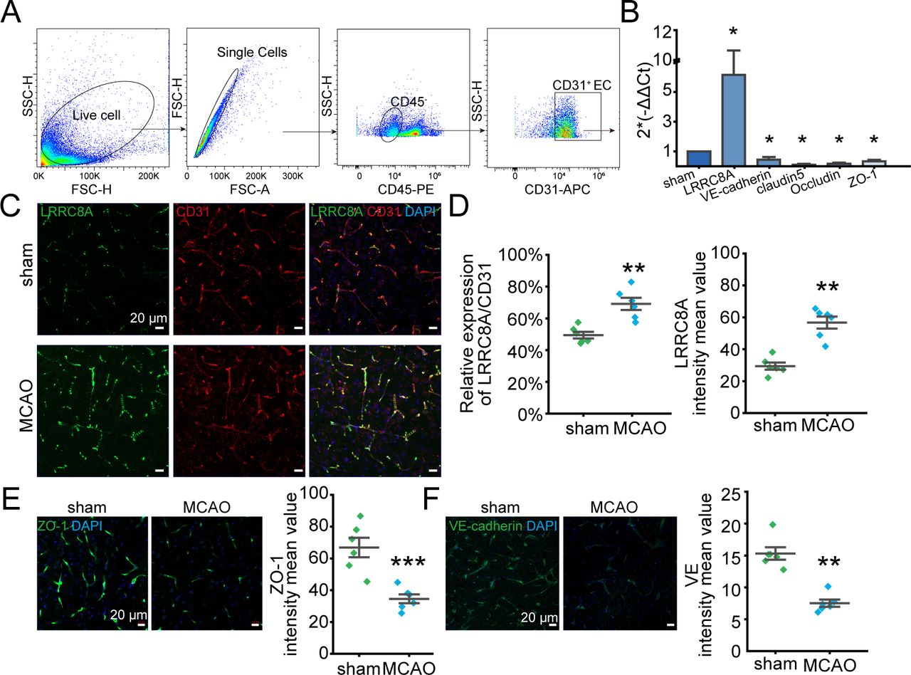
- Figure 2
LRRC8A expression was augmented in endothelial cells post-ischaemic stroke. Mice underwent MCAO surgery and sham operation 24 hours after reperfusion, LRRC8A expression in endothelial cells was tested. CD31 was used as the marker of endothelial cells. (A) CD45−CD31+ was used as the strategy of brain endothelial cell sorting. (B) mRNA levels of LRRC8A, VE-cadherin, claudin5, occludin and ZO-1 in sorted endothelial cells in sham and MCAO groups. Data were normalised to the sham group. Three mice were used for endothelial cell sorting each time and data represent three independent experiments. *P<0.05, two-sample t-test. (C) Representative images showed the co-expression of LRRC8A and CD31 in the sham and MCAO groups. (D) Analysis of fluorescence density. N=6 mice in each group. **P<0.01, two-sample t-test. (E, F) Left panel: representative images of ZO-1 (E) and VE-cadherin (F) in sham and MCAO groups. Right panel: quantification of ZO-1 and VE-cadherin expression. N=6 mice in each group. **P<0.01, two-sample t-test. LRRC8A, leucine-rich repeat-containing 8A; MCAO, middle cerebral artery occlusion.
- Figure 3
OGD augmented the VRCC current in both hBMVECs and mBMVECs that was mediated by LRRC8A. (A, B) Illustration of the experiment designs. (C) The hBMVECs were held at −60 mV and the currents were recorded using a hypertonic (420 mOsm) pipette solution. The representative traces of VRCC currents were inhibited by DCPIB (10 µM) at −60 mV preconditioning with OGD or under control conditions. (D, E) The representative current traces induced by step (D) or ramp (E) protocols were shown when the VRCC current in panel C reached the maximum at −60 mV preconditioning with OGD or under control conditions. (F) Summary data for the hBMVECs current densities recorded at −60 mV. N=12 cells in each group. *P<0.05, compared with hBMVECs control. (G) VRCC current was recorded with mBMVECs at −60 mV. (H, I) The representative current traces induced by step (H) or ramp (I) protocols were shown when the VRCC current in panel G reached the maximum at −60 mV. (J) Summary data for the mBMVECs current densities recorded at −60 mV. N=12 cells in each group. *P<0.05, compared with the mBMVECs group. (K) The representative VRCC current traces in the hBMVECs transfected with LRRC8A siRNA or scramble. (L, M) The representative current traces induced by step (L) or ramp (M) protocols were shown when the VRCC current in panel K reached the maximum at −60 mV. (N) Summary data for the hBMVECs VRCC current densities recorded at −60 mV transfected with LRRC8A siRNA or scramble. N=10 cells in each group. *P<0.05, compared with hBMVECs group. (O) The representative VRCC current traces in the mBMVECs transfected with LRRC8A siRNA or scramble. (P, Q) The representative current traces induced by step (P) or ramp (Q) protocols were shown when the VRCC current in panel O reached the maximum at −60 mV. (R) Summary data for the mBMVECs VRCC current densities recorded at −60 mV transfected with LRRC8A siRNA or scramble. N=10 cells in each group. *P<0.05, compared with mBMVECs group. All statistical analyses were performed using two-sample t-tests. hBMVEC, human brain microvascular endothelial cell; LRRC8A, leucine-rich repeat-containing 8A; mBMVEC, mouse brain microvascular endothelial cell; OGD, oxygen-glucose deprivation; VRCC, volume-regulated chloride channel.
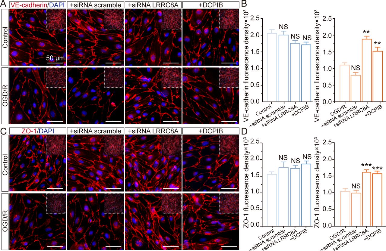
- Figure 4
Effect of LRRC8A modulation on ZO-1 and VE-cadherin expression in mBMVECs. mBMVECs were isolated and cultured for the BBB model in vitro. Four days later, a confluent monolayer with a tight junction between endothelial cells was produced. Then LRRC8A siRNA or scramble control was transfected. 48 hours later, cells were administrated with OGD exposure for 12 hours and followed by re-oxygenation (R) for 24 hours. Additionally, mBMVECs were pre-incubated with DCPIB (10 µM) for 30 min before OGD/R exposure. (A) and (C) Representative images (zoomed-in images of the inner part) showed the ZO-1 and VE-cadherin expression in different groups. (B) and (D) Quantification of ZO-1 and VE-cadherin expression in both the plasma membrane and cytoplasm from the inner part with the NIS-Elements AR Analysis software (V.5.01.00). Data represented the fluorescence intensity per unit area and were derived from four independent experiments. ***P<0.001, compared with the first column. **P<0.05, ***p<0.001, compared with the first column, one-way analysis of variance. BBB, blood-brain barrier; LRRC8A, leucine-rich repeat-containing 8A; mBMVEC, mouse brain microvascular endothelial cell; OGD, oxygen-glucose deprivation.
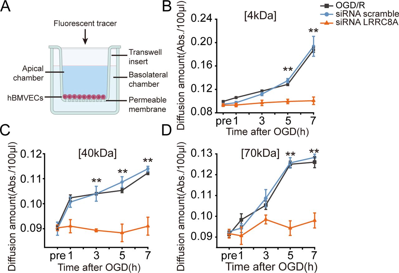
- Figure 5
LRRC8A knockout restored BBB integrity after OGD in vitro. (A) Schematic of the in vitro endothelial cell permeability assay. hBMVECs were seeded on the top insert and grown for 4 days to form a confluent monolayer. Then, hBMVECs were transfected with LRRC8A siRNA or scramble control for 48 hours, followed by OGD exposure for 12 hours. Cell permeability was evaluated at 1-, 3-, 5- and 7-hour post-OGD by the luminal to abluminal diffusion amount of three fluorescent tracers with different molecular weights. (B–D) Diffusion amounts of 4 kDa (B), 40 kDa (C) and 70 kDa (D) FITC-dextran at 1-, 3-, 5- and 7-hour post-OGD. Data represent six independent experiments. **P<0.01, compared with the scramble group, two-way analysis of variance. BBB, blood-brain barrier; hBMVEC, human brain microvascular endothelial cell; LRRC8A, leucine-rich repeat-containing 8A; OGD, oxygen-glucose deprivation.
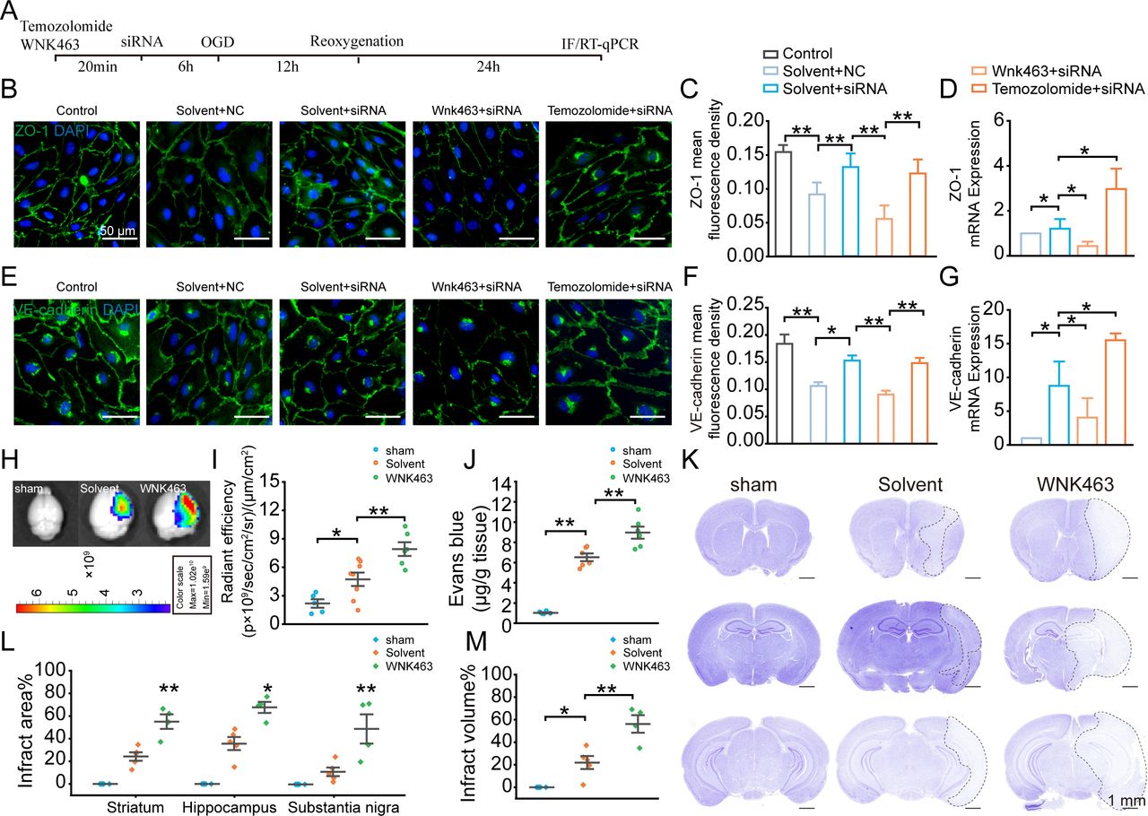
- Figure 6
WNK1-mediated LRRC8A-induced modulation of tight junctions. (A) Illustration of the experimental workflow. mBMVECs were incubated with 10 µM WNK463 (WNK1 inhibitor), 10 µM Temozolomide (WNK1 agonist) or solvent for 20 min, followed by transfection with siRNA LRRC8A or siRNA scramble. 48 hours later, the cells were subjected to OGD, followed by 24 hours of re-oxygenation. RNA was then extracted from the cells, or the cells were subjected to immunofluorescence staining to detect the expression levels of ZO-1 and VE-cadherin in each group. (B) Representative images of ZO-1 expression under different conditions. (C) Quantification of ZO-1 fluorescence density in both the plasma membrane and cytoplasm with ImageJ software (V.1.52a). Data represented the fluorescence intensity per unit area and were derived from three independent experiments. (D) Quantification of ZO-1 mRNA level. Data represented three independent experiments. *P<0.05, **p<0.01, one-way ANOVA. (E) Representative images of VE-cadherin expression under different conditions. (F) Quantification of VE-cadherin fluorescence density in both the plasma membrane and cytoplasm with ImageJ software (V.1.52a). Data represented the fluorescence intensity per unit area. (G) Quantification of VE-cadherin mRNA level. *P<0.05, **p<0.01, one-way ANOVA. (H–J) Endothelial cell-specific knockout of LRRC8A mice underwent sham and MCAO operation. Solvent and WNK463 were administrated before surgery. Representative images (H) and statistical analysis (I) demonstrated the extravasation of EB into the cortex. N=5 mice in sham group, n=8 mice in solvent group, n=6 mice in WNK463 group, *p<0.05, **p<0.01, one-way ANOVA. (J) Quantification of EB leakage with tissue homogenisation. N=6 mice in each group, **p<0.01, one-way ANOVA. (K) Representative Nissl-stained coronal sections 24 hours post-MCAO in endothelial cell-specific LRRC8A knockout mice. The white dotted lines indicated borders of the infarct zones. Sections were arranged from rostral to caudal at Bregma coordinates: 0.86 mm, −1.34 mm and −2.80 mm. (L) Quantification of the infarct area across coronal sections. N=5 mice in sham group, n=5 mice in solvent group, n=4 mice in WNK463 group, *p<0.05, **p<0.01, two-way ANOVA. (M) Quantification of total infarct volume. N=5 mice in sham group, n=5 mice in solvent group, n=4 mice in WNK463 group, *p<0.05, **p<0.01, one-way ANOVA. ANOVA, analysis of variance; BBB, blood-brain barrier; mBMVEC, mouse brain microvascular endothelial cell; LRRC8A, leucine-rich repeat-containing 8A; MCAO, middle cerebral artery occlusion; OGD, oxygen-glucose deprivation; WNK1, with-no-lysine kinase 1.
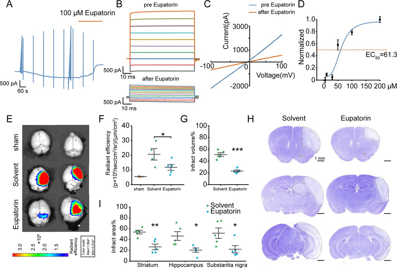
- Figure 7
Effect of Eupatorin on LRRC8A and brain injury post-ischaemic stroke. (A) hBMVECs were continuously held at −60 mV and stimulated with 1 s voltage ramps from −100 to +100 mV applied with a 60 s interval, 100 µM Eupatorin was applied when the current reached the maximum. (B) Effect of Eupatorin on the current–voltage (I/V) relationship, with voltage steps ranging from −100 to +100 mV in 20 mV increments. (C) The representative current traces induced by ramp protocols were shown before and Eupatorin application. (D) Dose–response curve of Eupatorin and LRRC8A current inhibition. N=6 cells for analysis. (E, F) Representative images (E) and statistical analysis (F) demonstrated the extravasation of EB into the cortex pretreated with Eupatorin or vehicle in wild-type mice. N=3 mice in sham group, n=4 mice in solvent group, n=5 in Eupatorin group, *p<0.05, one-way ANOVA. (G) Quantification of total infarct volume. N=5 mice in each group, ***p<0.001, two-sample t-test. (H) Representative images of Nissl staining. The white dotted lines indicated the borders of the infarct zones. Sections were arranged from rostral to caudal at Bregma coordinates: 0.86 mm, −1.34 mm and −2.80 mm. (I) Quantification of the infarct area across coronal sections. N=5 mice in each group, *p<0.05, **p<0.01, two-way ANOVA. ANOVA, analysis of variance; hBMVEC, human brain microvascular endothelial cell; LRRC8A, leucine-rich repeat-containing 8A.


Supplementary Materials
Supplementary data


