
Main menu

  • Online first
    • Online first
  • Current issue
    • Current issue
  • Archive
    • Archive
  • Submit a paper
    • Online submission site
    • Instructions for authors
  • About the journal
    • About the journal
    • Editorial board
    • Instructions for authors
    • FAQs
    • Chinese Stroke Association
  • Help
    • Contact us
    • Feedback form
    • Reprints
    • Permissions
    • Advertising
  • BMJ Journals

User menu

  • Login

Search

  • Advanced search
  • BMJ Journals
  • Login
  • Facebook
  • Twitter
Stroke and Vascular Neurology

Advanced Search

  • Online first
    • Online first
  • Current issue
    • Current issue
  • Archive
    • Archive
  • Submit a paper
    • Online submission site
    • Instructions for authors
  • About the journal
    • About the journal
    • Editorial board
    • Instructions for authors
    • FAQs
    • Chinese Stroke Association
  • Help
    • Contact us
    • Feedback form
    • Reprints
    • Permissions
    • Advertising
Open Access

Bone marrow-derived mesenchymal stem cell ameliorates post-stroke enterobacterial translocation through liver-gut axis

Xiaotao Su, Tiemei Li, Yuge Wang, Lei Wei, Banghao Jian, Xinmei Kang, Mengyan Hu, Chunyi Li, Shisi Wang, Danli Lu, Shishi Shen, Huipeng Huang, Yuxin Liu, Xiaohui Deng, Bingjun Zhang, Wei Cai, Zhengqi Lu
DOI: 10.1136/svn-2024-003494 Published 4 October 2024
Xiaotao Su
1Department of Neurology, Mental and Neurological Disease Research Center, The Third Affiliated Hospital of Sun Yat-Sen University, Guangzhou, Guangdong, China
2Center of Clinical Immunology, Mental and Neurological Disease Research Center, The Third Affiliated Hospital of Sun Yat-Sen University, Guangzhou, Guangdong, China
3Guangdong Provincial Key Laboratory of Brain Function and Disease, Guangzhou, Guangdong, China
  • Find this author on Google Scholar
  • Find this author on PubMed
  • Search for this author on this site
Tiemei Li
1Department of Neurology, Mental and Neurological Disease Research Center, The Third Affiliated Hospital of Sun Yat-Sen University, Guangzhou, Guangdong, China
  • Find this author on Google Scholar
  • Find this author on PubMed
  • Search for this author on this site
Yuge Wang
1Department of Neurology, Mental and Neurological Disease Research Center, The Third Affiliated Hospital of Sun Yat-Sen University, Guangzhou, Guangdong, China
  • Find this author on Google Scholar
  • Find this author on PubMed
  • Search for this author on this site
Lei Wei
1Department of Neurology, Mental and Neurological Disease Research Center, The Third Affiliated Hospital of Sun Yat-Sen University, Guangzhou, Guangdong, China
  • Find this author on Google Scholar
  • Find this author on PubMed
  • Search for this author on this site
Banghao Jian
1Department of Neurology, Mental and Neurological Disease Research Center, The Third Affiliated Hospital of Sun Yat-Sen University, Guangzhou, Guangdong, China
  • Find this author on Google Scholar
  • Find this author on PubMed
  • Search for this author on this site
Xinmei Kang
1Department of Neurology, Mental and Neurological Disease Research Center, The Third Affiliated Hospital of Sun Yat-Sen University, Guangzhou, Guangdong, China
  • Find this author on Google Scholar
  • Find this author on PubMed
  • Search for this author on this site
Mengyan Hu
1Department of Neurology, Mental and Neurological Disease Research Center, The Third Affiliated Hospital of Sun Yat-Sen University, Guangzhou, Guangdong, China
  • Find this author on Google Scholar
  • Find this author on PubMed
  • Search for this author on this site
Chunyi Li
1Department of Neurology, Mental and Neurological Disease Research Center, The Third Affiliated Hospital of Sun Yat-Sen University, Guangzhou, Guangdong, China
  • Find this author on Google Scholar
  • Find this author on PubMed
  • Search for this author on this site
Shisi Wang
1Department of Neurology, Mental and Neurological Disease Research Center, The Third Affiliated Hospital of Sun Yat-Sen University, Guangzhou, Guangdong, China
  • Find this author on Google Scholar
  • Find this author on PubMed
  • Search for this author on this site
Danli Lu
1Department of Neurology, Mental and Neurological Disease Research Center, The Third Affiliated Hospital of Sun Yat-Sen University, Guangzhou, Guangdong, China
  • Find this author on Google Scholar
  • Find this author on PubMed
  • Search for this author on this site
Shishi Shen
1Department of Neurology, Mental and Neurological Disease Research Center, The Third Affiliated Hospital of Sun Yat-Sen University, Guangzhou, Guangdong, China
  • Find this author on Google Scholar
  • Find this author on PubMed
  • Search for this author on this site
Huipeng Huang
1Department of Neurology, Mental and Neurological Disease Research Center, The Third Affiliated Hospital of Sun Yat-Sen University, Guangzhou, Guangdong, China
  • Find this author on Google Scholar
  • Find this author on PubMed
  • Search for this author on this site
Yuxin Liu
1Department of Neurology, Mental and Neurological Disease Research Center, The Third Affiliated Hospital of Sun Yat-Sen University, Guangzhou, Guangdong, China
  • Find this author on Google Scholar
  • Find this author on PubMed
  • Search for this author on this site
Xiaohui Deng
1Department of Neurology, Mental and Neurological Disease Research Center, The Third Affiliated Hospital of Sun Yat-Sen University, Guangzhou, Guangdong, China
  • Find this author on Google Scholar
  • Find this author on PubMed
  • Search for this author on this site
Bingjun Zhang
1Department of Neurology, Mental and Neurological Disease Research Center, The Third Affiliated Hospital of Sun Yat-Sen University, Guangzhou, Guangdong, China
  • Find this author on Google Scholar
  • Find this author on PubMed
  • Search for this author on this site
Wei Cai
1Department of Neurology, Mental and Neurological Disease Research Center, The Third Affiliated Hospital of Sun Yat-Sen University, Guangzhou, Guangdong, China
2Center of Clinical Immunology, Mental and Neurological Disease Research Center, The Third Affiliated Hospital of Sun Yat-Sen University, Guangzhou, Guangdong, China
3Guangdong Provincial Key Laboratory of Brain Function and Disease, Guangzhou, Guangdong, China
  • Find this author on Google Scholar
  • Find this author on PubMed
  • Search for this author on this site
  • ORCID record for Wei Cai
Zhengqi Lu
1Department of Neurology, Mental and Neurological Disease Research Center, The Third Affiliated Hospital of Sun Yat-Sen University, Guangzhou, Guangdong, China
  • Find this author on Google Scholar
  • Find this author on PubMed
  • Search for this author on this site
  • ORCID record for Zhengqi Lu
  • Article
  • Figures & Data
  • eLetters
  • Info & Metrics
  • PDF
Loading

Article Figures & Data

Figures

  • Supplementary Materials
  • Additional Files
  • Figure 1
    • Download figure
    • Open in new tab
    • Download powerpoint
    Figure 1

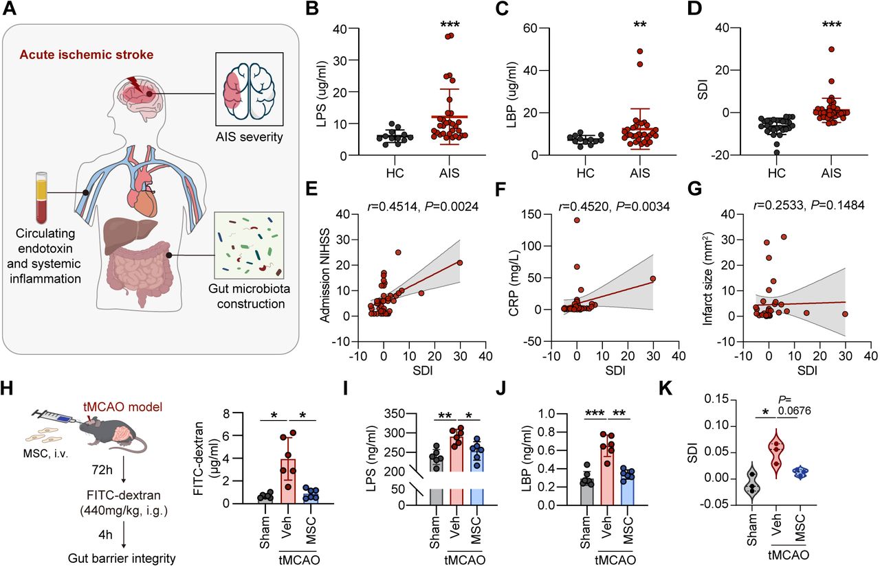
    BM-MSC transfer ameliorates post-stroke enterobacterial translocation. (A) Experimental design of gut bacteria translocation in patients with AIS. Serum LPS (B) and LBP (C) level of patients with AIS (N=31) and HC (N=12) assessed by ELISA. *p<0.05, **p<0.01; by Welch’s t-test (mean±SD). (D) Comparison of SDI in patients with AIS (N=43) and HC (N=28), ***p<0.001, by Welch’s t-test (mean±SD). SDI of patients who had a stroke correlating with the admission NIHSS score (E), CRP level (F) and infarct size (G) in patients with AIS, by Spearman rank correlation test. (H–K) Wild-type C57/BL6 mice were subjected to 60 min of tMCAO and transferred with a single dose of BM-MSC (2×10⁶ cells per mice, intravenous) or an equal volume of vehicle (Veh, intravenous) right after reperfusion. Serum and faecal samples were collected at 3 days after tMCAO. (H) Schematic diagram of the gut barrier integrity assay and the results of fluorescence quantification. N=6 in each group, *p<0.05, by Welch ANOVA (mean±SD). Serum LPS (I) and LBP (J) level of sham and stroke mice. N=6 mice in each group, *p<0.05, **p<0.01, ***p<0.001, by Welch ANOVA (mean±SD). (K) Comparison of SDI in sham and stroke mice. N=3 mice in each group, *p<0.05, by Welch ANOVA (mean±SD). AIS, acute ischaemic stroke; ANOVA, analysis of variance; BM-MSC, bone marrow-derived mesenchymal stem cell; CRP, C reactive protein; FITC, fluorescein isothiocyanate; HC, healthy control; LBP, lipopolysaccharides binding protein; LPS, lipopolysaccharides; NIHSS, National Institute of Health stroke scale; SDI, Stroke Dysbiosis Index; tMCAO, transient middle cerebral artery occlusion.

  • Figure 2
    • Download figure
    • Open in new tab
    • Download powerpoint
    Figure 2

    BM-MSC transfer does not modulate gut structure. (A) Weight loss (d3–d1) in mice of sham, tMCAO+Veh and tMCAO+MSC. N=6 mice in each group. ***p<0.001, compared with sham group. Not significant, by one-way ANOVA (mean±SD). (B) Representative picture of colon in sham, tMCAO+Veh and tMCAO+MSC mice and the quantification of colon length. N=6 mice in each group. Not significant, by one-way ANOVA (mean±SD). (C) Colon H&E stained and statistics of the histological score, crypt length and width in sham, tMCAO+Veh and tMCAO+MSC mice. N=6 mice in each group. Not significant, by one-way ANOVA (mean±SD). (D) Heatmap of tight junction-related genes of colon tissue between Veh and MSC after 3 days of tMCAO according to bulk RNA sequencing (RNA-seq), N=3 mice in each group. (E–F) Representative images of ZO1(E) and CLDN4 (F) of colon tissue in mice of in sham, tMCAO+Veh and tMCAO+MSC groups. N=3 mice in each group. ANOVA, analysis of variance; BM-MSC, bone marrow-derived mesenchymal stem cell; CLDN4, Claudin 4; tMCAO, transient middle cerebral artery occlusion; ZO1, zona occludens 1.

  • Figure 3
    • Download figure
    • Open in new tab
    • Download powerpoint
    Figure 3

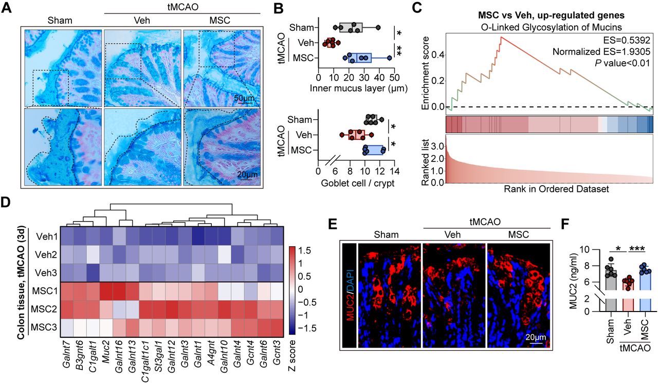
    Colon mucus layer is attenuated after stroke which is preserved by BM-MSC transferred. (A–B) Alcian stain of colon tissue of sham, tMCAO+Veh and tMCAO+MSC mice. N=6 mice in each group. (A) Representative image of the mucus layer. (B) Corresponding quantitation of mucus layer thickness and number of goblet cells. Data were analysed using one-way ANOVA (mean±SD). *p<0.05, **p<0.01. (C–D) Analysis of sequencing results of colon tissue between tMCAO+Veh and tMCAO+MSC group. N=3 mice in each group. (C) GSEA showed that the upregulated genes in the MSC-treated group were enriched in the O-linked glycosylation of mucins pathway and the specific gene expression of this pathway was shown in the heatmap (D). (E) Immunostaining showed increased expression of MUC2 in the colon of BM-MSC-transferred mice. N=3 mice in each group. (F) Expression of colon MUC2 was quantified by ELISA. N=6 mice in each group. *p<0.05, ***p<0.001, by one-way ANOVA (mean±SD). ANOVA, analysis of variance; BM-MSC, bone marrow-derived mesenchymal stem cell; GSEA, gene set enrichment analysis; MUC2, Mucin-2; tMCAO, transient middle cerebral artery occlusion.

  • Figure 4
    • Download figure
    • Open in new tab
    • Download powerpoint
    Figure 4

    BM-MSC improves gut microbiota construction through preserving the colon mucus layer. (A–F) Faecal samples of mice at day 3 after tMCAO were collected and performed the 16S rRNA sequencing for the following analysis. N=3 mice in each group. (A) PCoA of the gut microbiome composition on the genus level. (B) Cluster analysis was performed using the unweighted pair group method with arithmetic mean (UPGMA) based on the weighted UniFrac distances to compare the community composition similarity of samples. (C) Characteristics of gut microbiota in the groups were evaluated with LEfSe. Bacterial populations with LDA score >3.5 were displayed. Relative abundance of gut bacterial phylum (D) and species (E) level in each group at day 3 after tMCAO. (F) Cladogram based on LEfSe analysis depicted the phylogenetic tree of the taxa enriched in microbiota from tMCAO mice received BM-MSC. (G–I) Gut microbiota was depleted with pretreatment of broad-spectrum antibiotics (ABX) for 28 days before tMCAO. (G) Experimental design. (H) Representative images of Alcian blue staining and statistics of mucus layer thickness showed that the repairing effect of BM-MSC on the mucus layer was inhibited by dexamethasone. N=4 mice of each group in the experiment measured the mucus layer, **p<0.01, by Student’s t-test (mean±SD). N=7 mice in each group in the experiment measured the number of goblet cells. ***p<0.001, by Welch’s t-test (mean±SD). (I) Gut barrier integrity was assessed as the results of fluorescence quantification of FITC-dextran. N=5 mice in each group, ***p<0.001, by Student’s t-test (mean±SD). BM-MSC, bone marrow-derived mesenchymal stem cell; FITC, fluorescein isothiocyanate; LDA, linear discriminant analysis; LEfSe, linear discriminant analysis effect size; PCoA, principal coordinate analysis; rRNA, ribosomal RNA; tMCAO, transient middle cerebral artery occlusion.

  • Figure 5
    • Download figure
    • Open in new tab
    • Download powerpoint
    Figure 5

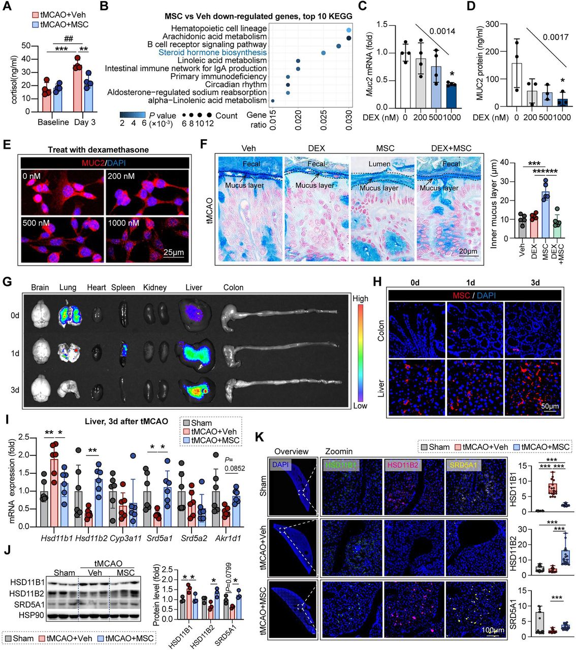
    BM-MSC preserves gut barrier integrity through liver-gut axis. (A) Changes in plasma cortisol concentrations after stroke in mice. N=4 mice in each group. **p<0.01, ***p<0.001, comparisons were made between the two groups labelled. ##p<0.01 compared with baseline cortisol level in the whole experiment, by two-way ANOVA (mean±SD). (B) The differentially expressed genes down-regulated in the colons of BM-MSC treated group were enriched in the steroid hormone biosynthesis pathway. (C–E) To explore the inhibitory effect of glucocorticoid on the mucus layer, goblet cells were treated with a concentration gradient of dexamethasone for 18 hours. The transcription and translation of MUC2 were assessed by QPCR (C) and ELISA (D), respectively. Experiments were repeated for 3–4 times. *p<0.05, compared with the control group (0 nM), by one-way ANOVA (mean±SD). The slope of Muc2 mRNA and MUC2 protein expression line treated with the concentration gradient dexamethasone were also calculated by simple linear regression. (E) Expression of MUC2 (red) of goblet cells after concentration gradients of dexamethasone treatment was assessed by immunofluorescence staining. Experiments were repeated for three times. In addition, the dexamethasone treatment at these concentrations did not accelerate apoptosis in goblet cells. Data were shown in online supplemental figure 5A. (F) Representative images of Alcian blue staining and statistics of mucus layer thickness showed that the repairing effect of BM-MSC on the mucus layer was inhibited by dexamethasone. N=5 in each group. ***p<0.001, by one-way ANOVA (mean±SD). Additional data for dexamethasone treatment under these subgroups were presented in online supplemental figure B–D. (G) In vivo imaging of major organs from tMCAO mice at 0 day, 1 day and 3 days after intravenously injection of DiR-labelled BM-MSC (2×10⁶ cells per mouse). N=3 mice in each group. (H) Infiltration of WGA-labelled BM-MSC of the liver and colon at 0 day, 1 day and 3 days after intravenously injection were tracked by immunofluorescence. N=3 mice in each group. (I–K) Liver tissue of stroke mice was isolated at 3 days after tMCAO and used for the following experiments. The expression of enzymes that regulated the level and effects of glucocorticoid were detected by QPCR array (I). N=6 mice in each group. *p<0.05, **p<0.01, by one-way ANOVA (mean±SD). The change of HSD11B1, HSD11B2 and SRD5A1 were further verified by Western blot (J) and immunostaining (K). N=3 mice in each group. *p<0.05, ***p<0.001, by one-way ANOVA (mean±SD). ANOVA, analysis of variance; BM-MSC, bone marrow-derived mesenchymal stem cell; DAPI, 4‘,6-diamidino-2-phenylindole; DEX, dexamethasone; IgA, immunoglobulin A; KEGG, Kyoto Encyclopedia of Genes and Genomes; mRNA, messenger RNA; MUC2, Mucin-2; QPCR, quantitative PCR; tMCAO, transient middle cerebral artery occlusion.

  • Figure 6
    • Download figure
    • Open in new tab
    • Download powerpoint
    Figure 6

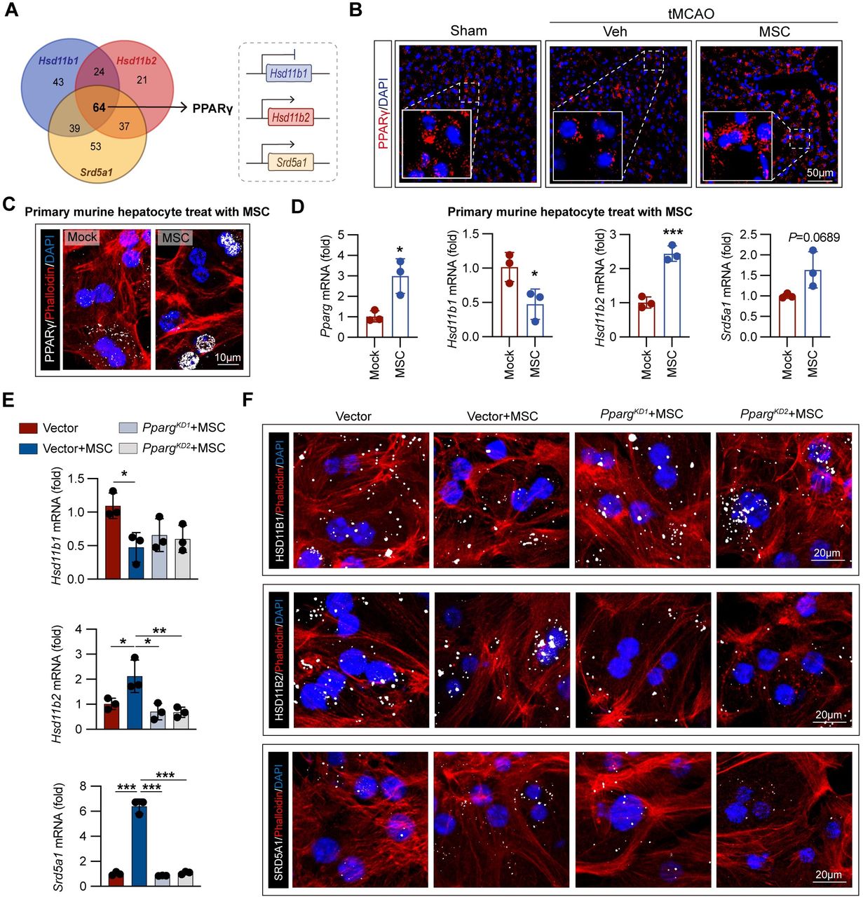
    BM-MSC accelerates GC inactivation through enhancing PPARγ signalling in hepatocytes. (A) Venn diagram showed the intersection of TFs that modulated the transcription of Hsd11b1, Hsd11b2 and Srd5a1. Among the 64 TFs intersected, PPARγ could simultaneously downregulated the transcription of Hsd11b1 and upregulate the transcription of Hsd11b2 and Srd5a1. Data for the analysis were obtained from the signalling pathways project (SPP, http://signalingpathways.org/). (B) Liver sections were subjected to immunostaining of PPARγ (red) and DAPI (blue) at 3 days after tMCAO. Experiments were repeated for three times. (C–D) Murine primary hepatocytes were co-cultured with BM-MSC (hepatocytes:BM-MSC=5:1) for 18 hours. (C) Immunofluorescence images showed enhanced nuclear localisation of PPARγ (grey) after co-culture of murine primary hepatocytes with BM-MSC. Experiments were repeated for three times. (D) The mRNA level of Pparg, Hsd11b1, Hsd11b2 and Srd5a1 in murine primary hepatocytes were assessed with QPCR. Experiments were repeated for three times. *p<0.05, ***p<0.001, by one-way ANOVA (mean±SD). (E–F) Pparg was knocked-down in murine primary hepatocytes by lentivirus-shRNA (PpargKD). Murine primary hepatocytes transfected with vector plasmid were set as control. The effect of Pparg in the promotion of glucocorticoid metabolism in hepatocytes by BM-MSC was verified by QPCR (E) and immunofluorescence (F). Experiments were repeated three times. *p<0.05, **p<0.01, ***p<0.001, by one-way ANOVA (mean±SD). ANOVA, analysis of variance; BM-MSC, bone marrow-derived mesenchymal stem cell; DAPI, 4‘,6-diamidino-2-phenylindole; mRNA, messenger RNA; PPARγ, peroxisome proliferator-activated receptor γ; QPCR, quantitative PCR; shRNA, short hairpin RNA; TFs, transcriptional factors; tMCAO, transient middle cerebral artery occlusion.

Supplementary Materials

  • Figures
  • Additional Files
  • Supplementary data

    [svn-2024-003494supp001.pdf]

Additional Files

  • Figures
  • Supplementary Materials
  • Supplementary Data

    This web only file has been produced by the BMJ Publishing Group from an electronic file supplied by the author(s) and has not been edited for content.

    • Data supplement 1
PreviousNext
Back to top
Vol 10 Issue 1 Table of Contents
Stroke and Vascular Neurology: 10 (1)
  • Table of Contents
  • Table of Contents (PDF)
  • About the Cover
  • Index by author
  • Ed Board (PDF)
Email

Thank you for your interest in spreading the word on Stroke and Vascular Neurology.

NOTE: We only request your email address so that the person you are recommending the page to knows that you wanted them to see it, and that it is not junk mail. We do not capture any email address.

Enter multiple addresses on separate lines or separate them with commas.
Bone marrow-derived mesenchymal stem cell ameliorates post-stroke enterobacterial translocation through liver-gut axis
(Your Name) has sent you a message from Stroke and Vascular Neurology
(Your Name) thought you would like to see the Stroke and Vascular Neurology web site.
CAPTCHA
This question is for testing whether or not you are a human visitor and to prevent automated spam submissions.
Print
Alerts
Sign In to Email Alerts with your Email Address
Citation Tools
Bone marrow-derived mesenchymal stem cell ameliorates post-stroke enterobacterial translocation through liver-gut axis
Xiaotao Su, Tiemei Li, Yuge Wang, Lei Wei, Banghao Jian, Xinmei Kang, Mengyan Hu, Chunyi Li, Shisi Wang, Danli Lu, Shishi Shen, Huipeng Huang, Yuxin Liu, Xiaohui Deng, Bingjun Zhang, Wei Cai, Zhengqi Lu
Stroke and Vascular Neurology Oct 2024, svn-2024-003494; DOI: 10.1136/svn-2024-003494

Citation Manager Formats

  • BibTeX
  • Bookends
  • EasyBib
  • EndNote (tagged)
  • EndNote 8 (xml)
  • Medlars
  • Mendeley
  • Papers
  • RefWorks Tagged
  • Ref Manager
  • RIS
  • Zotero
Cite This
  • APA
  • Chicago
  • Endnote
  • MLA
Loading
Bone marrow-derived mesenchymal stem cell ameliorates post-stroke enterobacterial translocation through liver-gut axis
Xiaotao Su, Tiemei Li, Yuge Wang, Lei Wei, Banghao Jian, Xinmei Kang, Mengyan Hu, Chunyi Li, Shisi Wang, Danli Lu, Shishi Shen, Huipeng Huang, Yuxin Liu, Xiaohui Deng, Bingjun Zhang, Wei Cai, Zhengqi Lu
Stroke and Vascular Neurology Oct 2024, svn-2024-003494; DOI: 10.1136/svn-2024-003494
Download PDF

Share
Bone marrow-derived mesenchymal stem cell ameliorates post-stroke enterobacterial translocation through liver-gut axis
Xiaotao Su, Tiemei Li, Yuge Wang, Lei Wei, Banghao Jian, Xinmei Kang, Mengyan Hu, Chunyi Li, Shisi Wang, Danli Lu, Shishi Shen, Huipeng Huang, Yuxin Liu, Xiaohui Deng, Bingjun Zhang, Wei Cai, Zhengqi Lu
Stroke and Vascular Neurology Oct 2024, svn-2024-003494; DOI: 10.1136/svn-2024-003494
Reddit logo Twitter logo Facebook logo Mendeley logo
Respond to this article
  • Tweet Widget
  • Facebook Like
  • Google Plus One
  • Article
    • Abstract
    • Introduction
    • Materials and methods
    • Results
    • Discussion
    • Data availability statement
    • Ethics statements
    • Acknowledgments
    • Footnotes
    • References
  • Figures & Data
  • eLetters
  • Info & Metrics
  • PDF

Related Articles

Cited By...

More in this TOC Section

  • Learning curve and embolisation strategy in single-stage surgery combined embolisation and microsurgery for brain arteriovenous malformations: results from a nationwide multicentre prospective registry study
  • Stepwise improvement in intracerebral haematoma expansion prediction with advanced imaging: a comprehensive comparison of existing scores
  • Thrombus iodine-based perviousness is associated with recanalisation and functional outcomes in endovascular thrombectomy
Show more Original research

Similar Articles


CONTENT

  • Latest content
  • Current issue
  • Archive
  • eLetters
  • Sign up for email alerts
  • RSS

JOURNAL

  • About the journal
  • Editorial board
  • Recommend to librarian
  • Chinese Stroke Association

AUTHORS

  • Instructions for authors
  • Submit a paper
  • Track your article
  • Open Access at BMJ

HELP

  • Contact us
  • Reprints
  • Permissions
  • Advertising
  • Feedback form

© 2025 Chinese Stroke Association