
Main menu

  • Online first
    • Online first
  • Current issue
    • Current issue
  • Archive
    • Archive
  • Submit a paper
    • Online submission site
    • Instructions for authors
  • About the journal
    • About the journal
    • Editorial board
    • Instructions for authors
    • FAQs
    • Chinese Stroke Association
  • Help
    • Contact us
    • Feedback form
    • Reprints
    • Permissions
    • Advertising
  • BMJ Journals

User menu

  • Login

Search

  • Advanced search
  • BMJ Journals
  • Login
  • Facebook
  • Twitter
Stroke and Vascular Neurology

Advanced Search

  • Online first
    • Online first
  • Current issue
    • Current issue
  • Archive
    • Archive
  • Submit a paper
    • Online submission site
    • Instructions for authors
  • About the journal
    • About the journal
    • Editorial board
    • Instructions for authors
    • FAQs
    • Chinese Stroke Association
  • Help
    • Contact us
    • Feedback form
    • Reprints
    • Permissions
    • Advertising
Open Access

Cerebral small vessel disease modifies outcomes after minimally invasive surgery for intracerebral haemorrhage

Yunke Li, Sung-Min Cho, Radhika Avadhani, Hassan Ali, Yi Hao, Santosh B Murthy, Joshua N Goldstein, Fan Xia, Xin Hu, Natalie L Ullman, Issam Awad, Daniel Hanley, Wendy C Ziai
DOI: 10.1136/svn-2023-002463 Published 10 November 2023
Yunke Li
1The George Institute for Global Health, Beijing, China
  • Find this author on Google Scholar
  • Find this author on PubMed
  • Search for this author on this site
Sung-Min Cho
2Department of Neurology, Division of Neurocritical Care, Johns Hopkins Medical Institutions, Baltimore, Maryland, USA
  • Find this author on Google Scholar
  • Find this author on PubMed
  • Search for this author on this site
Radhika Avadhani
3Department of Neurology, Division of Brain Injury Outcomes, Johns Hopkins Medical Institutions Campus, Baltimore, Maryland, USA
  • Find this author on Google Scholar
  • Find this author on PubMed
  • Search for this author on this site
Hassan Ali
3Department of Neurology, Division of Brain Injury Outcomes, Johns Hopkins Medical Institutions Campus, Baltimore, Maryland, USA
  • Find this author on Google Scholar
  • Find this author on PubMed
  • Search for this author on this site
Yi Hao
3Department of Neurology, Division of Brain Injury Outcomes, Johns Hopkins Medical Institutions Campus, Baltimore, Maryland, USA
  • Find this author on Google Scholar
  • Find this author on PubMed
  • Search for this author on this site
Santosh B Murthy
4Department of Neurology, Weill Cornell Medical College, New York, New York, USA
  • Find this author on Google Scholar
  • Find this author on PubMed
  • Search for this author on this site
Joshua N Goldstein
5Department of Emergency Medicine, Massachusetts General Hospital, Boston, Massachusetts, USA
  • Find this author on Google Scholar
  • Find this author on PubMed
  • Search for this author on this site
Fan Xia
6Department of Neurosurgery, West China Hospital of Medicine, Chengdu, Sichuan, China
  • Find this author on Google Scholar
  • Find this author on PubMed
  • Search for this author on this site
Xin Hu
6Department of Neurosurgery, West China Hospital of Medicine, Chengdu, Sichuan, China
  • Find this author on Google Scholar
  • Find this author on PubMed
  • Search for this author on this site
Natalie L Ullman
3Department of Neurology, Division of Brain Injury Outcomes, Johns Hopkins Medical Institutions Campus, Baltimore, Maryland, USA
  • Find this author on Google Scholar
  • Find this author on PubMed
  • Search for this author on this site
Issam Awad
7Department of Neurosurgery, University of Chicago Pritzker School of Medicine, Chicago, Illinois, USA
  • Find this author on Google Scholar
  • Find this author on PubMed
  • Search for this author on this site
Daniel Hanley
3Department of Neurology, Division of Brain Injury Outcomes, Johns Hopkins Medical Institutions Campus, Baltimore, Maryland, USA
  • Find this author on Google Scholar
  • Find this author on PubMed
  • Search for this author on this site
Wendy C Ziai
2Department of Neurology, Division of Neurocritical Care, Johns Hopkins Medical Institutions, Baltimore, Maryland, USA
3Department of Neurology, Division of Brain Injury Outcomes, Johns Hopkins Medical Institutions Campus, Baltimore, Maryland, USA
  • Find this author on Google Scholar
  • Find this author on PubMed
  • Search for this author on this site
  • ORCID record for Wendy C Ziai
  • Article
  • Figures & Data
  • eLetters
  • Info & Metrics
  • PDF
Loading

Article Figures & Data
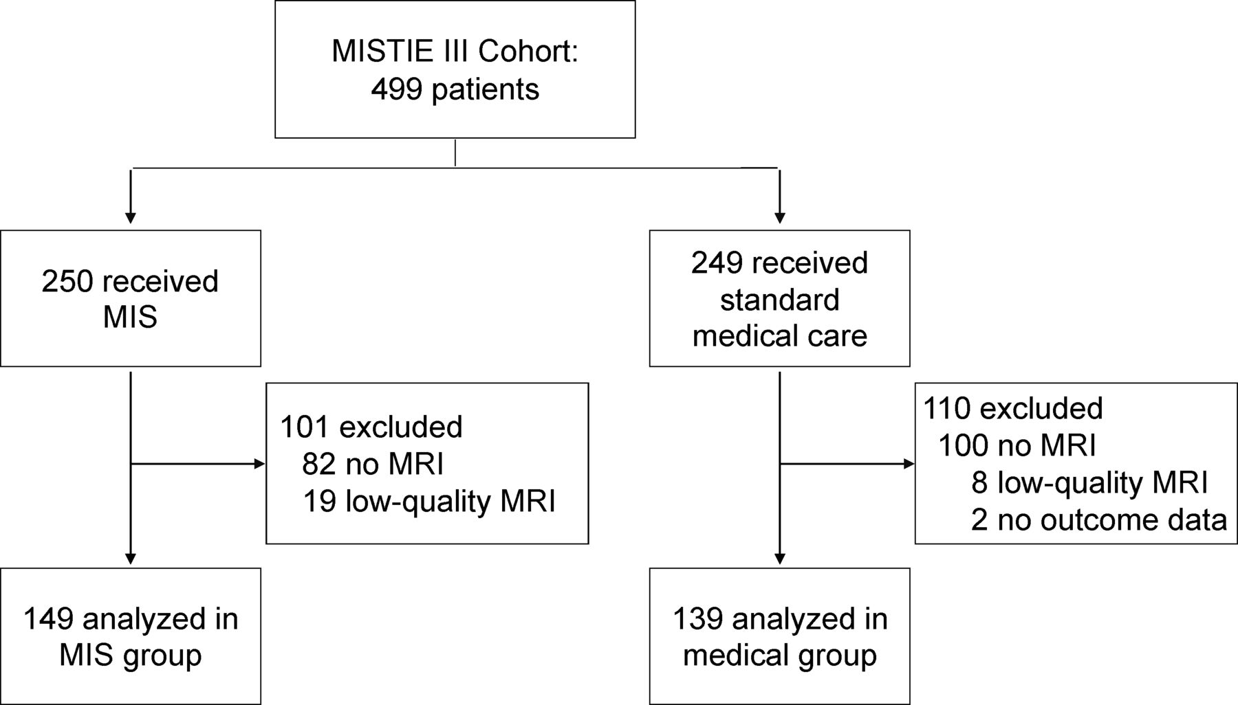
Figures

  • Tables
  • Supplementary Materials
  • Additional Files
  • Figure 1
    • Download figure
    • Open in new tab
    • Download powerpoint
    Figure 1

    Flowchart of patients from MISTIE III trial. MIS, minimally invasive surgery; MISTIE III, phase III minimally invasive surgery plus alteplase for intracerebral haemorrhage evacuation.
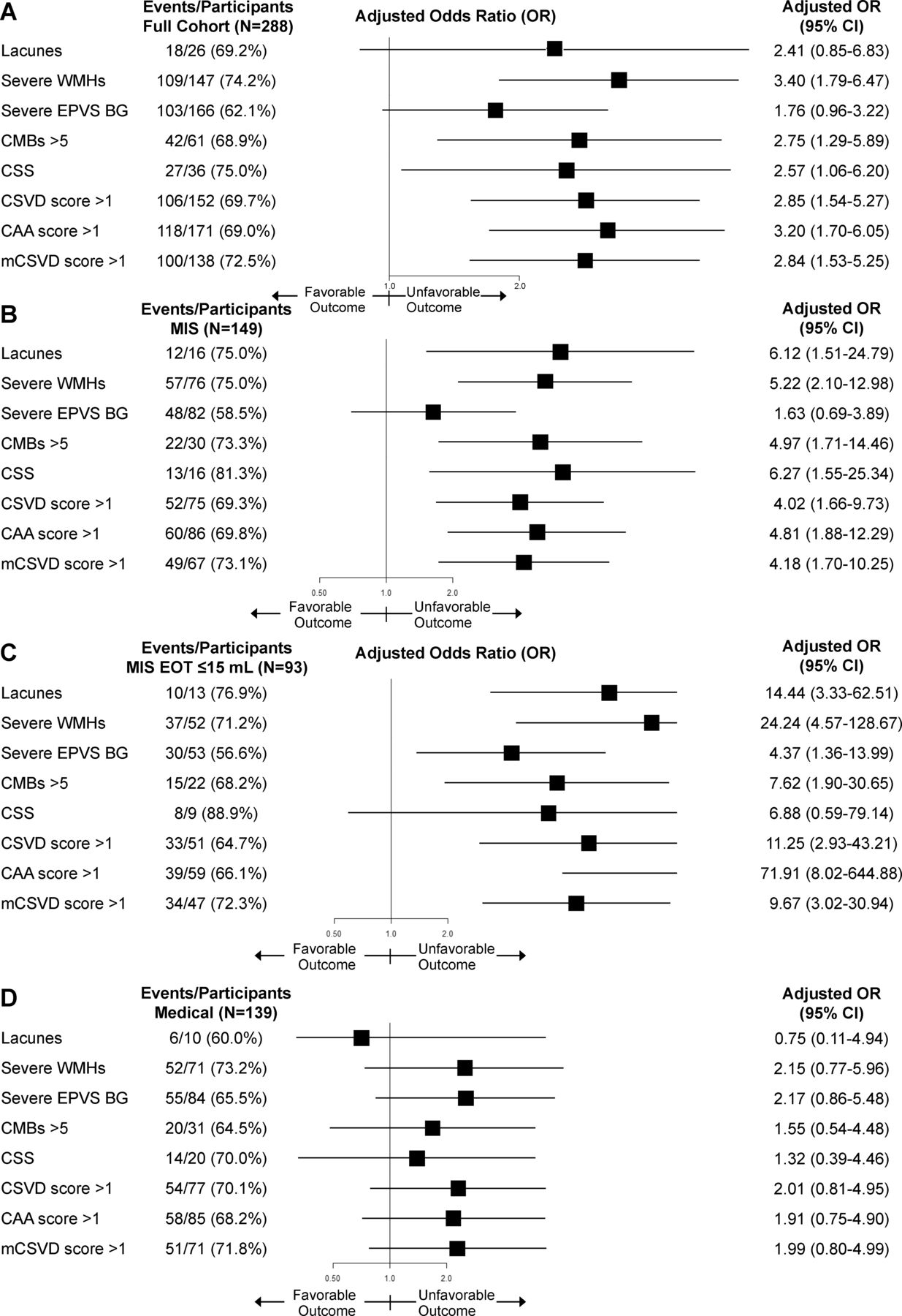
  • Figure 2
    • Download figure
    • Open in new tab
    • Download powerpoint
    Figure 2

    Prespecified adjusted subgroup analyses of odds of poor functional outcome at 1 year by brain MRI features for (A) full cohort, (B) MIS, (C) EOT volume ≤15 mL and (D) standard medical care (medical) group. All analyses were adjusted for age, admission GCS, diagnostic ICH volume, IVH present at diagnosis, lobar ICH location, diabetes and admission systolic blood pressure. BG, basal ganglia; CAA, cerebral amyloid angiopathy; CMB, cerebral microbleed; CSS, cortical superficial siderosis; CSVD, cerebral small vessel disease; EOT, end-of-treatment; EPVS, enlarged perivascular spaces; GCS, Glasgow Coma Scale; ICH, intracerebral haemorrhage; IVH, intraventricular haemorrhage; mCSVD, modified CSVD; MIS, minimally invasive surgery.

  • Figure 3
    • Download figure
    • Open in new tab
    • Download powerpoint
    Figure 3

    Pre-specified adjusted subgroup analysis of the odds of poor functional outcome at 1 year by brain MRI features comparing (A) MIS versus SMC, (B) EOT volume≤15 mL versus SMC and (C) EOT volume≤15 mL versus EOT volume>15 mL. All analyses were adjusted for age, admission GCS, diagnostic ICH volume, IVH present at diagnosis, lobar ICH location, diabetes and admission systolic blood pressure. BG, basal ganglia; CAA, cerebral amyloid angiopathy; CMB, cerebral microbleed; CSS, cortical superficial siderosis; CSVD, cerebral small vessel disease; EOT, end-of-treatment; EPVS, enlarged perivascular spaces; GCS, Glasgow Coma Scale; ICH, intracerebral haemorrhage; IVH, intraventricular haemorrhage; mCSVD, modified CSVD; MIS, minimally invasive surgery.

  • Figure 4
    • Download figure
    • Open in new tab
    • Download powerpoint
    Figure 4

    mRS score distribution by CAA score 0–1 versus 2–6 in (A) full MRI cohort (n=288), (B) MIS (n=149) versus medical (n=139) groups and (C) EOT volume≤15 mL (n=94) versus>15 mL (n=194). mRS scores range from 0 (no disability) to 6 (death). P values are for odds of mRS 0–3 versus 4–6 for CAA score 0–1 versus 2–6 in analyses adjusted for age, admission GCS, diagnostic ICH volume, IVH present at diagnosis, lobar ICH location, diabetes and admission systolic blood pressure. CAA, cerebral amyloid angiopathy; CSVD, cerebral small vessel disease; EOT, end-of-treatment; GCS, Glasgow Coma Scale; ICH, intracerebral haemorrhage; IVH, intraventricular haemorrhage; mCSVD, modified CSVD; MIS, minimally invasive surgery; mRS modified Rankin Score.

Tables

  • Figures
  • Supplementary Materials
  • Additional Files
  • Table 1

    Baseline characteristics of participants in the MRI substudy

    Standard medical care (n=139)MISTIE (n=149)EOT volume ≤15 mL (n=94)
    Sex, number (%)
     Male85 (61)89 (60)56 (60)
     Female56 (39)60 (40)38 (40)
    Age, median (IQR), years62 (52–71)62 (52–68)63 (52–69)
    Hypertension, number (%)137 (99)143 (96)92 (98)
    Diabetes, number (%)45 (32)45 (30)27 (29)
    Coronary artery disease, number (%)17 (12)23 (17)13 (14)
    Anticoagulant use, number (%)4 (3)15 (10)4 (4)
    Antiplatelet use, number (%)52 (37)44 (30)28 (30)
    Current smoker, number (%)19 (14)37 (25)25 (27)
    Alcohol abuse, number (%)12 (9)26 (17)13 (14)
    Baseline CT and clinical factors
     Diagnostic ICH volume, median (IQR), mL40.5 (30.8–54.9)42.2 (30.7–51.3)36.8 (29.3–49.6)
     Diagnostic IVH volume, median (IQR), mL0 (0–1.8)0 (0–1.6)0 (0–1.1)
     IVH present at diagnosis, number (%)57 (41)57 (38)32 (34)
     Lobar haematoma, number (%)58 (42)54 (36)30 (32)
     GCS at randomisation, median (IQR)10 (8–13)10 (8–13)10 (8–13)
     Systolic blood pressure, median (IQR), mm Hg180 (160–202)181 (155–208)180 (149–213)
     Diastolic blood pressure, median (IQR), mm Hg98 (84–119)98 (84–115)98 (84–115)
     Mean arterial pressure at first 24 hours, median (IQR)92 (83–98)92 (86–97)93 (86–98)
     Surgical group, number (%)0149 (100)91 (97)
     EOT CT ICH volume ≤15 mL, number (%)3 (2)91 (61)94 (100)
    • EOT, end-of-treatment; GCS, glasgow coma scale; ICH, intracerebral haemorrhage; INR, international normalised ratio; IVH, intraventricular haemorrhage; MISTIE, minimally invasive surgery plus alteplase for intracerebral haemorrhage evacuation; PTT, partial thromboplastin time.

  • Table 2

    Comparison of cerebral small vessel disease (CSVD) features and CSVD burden scores between outcome groups

    Full cohort, number (%) (n=288)
    Entire cohort
    (n=288)
    Favourable outcome
    (n=127)
    Unfavourable outcome (n=161)P value
    Presence of lacunes26 (9)8 (6)18 (11)0.21
    Lobar lacunes9 (3)4 (3)5 (3)1.00
    Deep lacunes17 (6)4 (3)13 (8)0.13
    Presence of CMBs118 (41)45 (35)73 (45)0.09
    Deep CMBs0.06
     0214 (74)102 (80)112 (70)
     131 (11)16 (13)15 (9)
     2–420 (7)6 (5)14 (9)
     5–1022 (8)7 (6)15 (9)
     >105 (2)0 (0)5 (3)
    Lobar CMBs0.16
     0196 (68)92 (72)104 (65)
     131 (11)16 (13)15 (9)
     2–420 (7)6 (5)14 (9)
     5–1030 (10)11 (9)19 (12)
     >1011 (4)2 (2)9 (6)
    Total CMBs0.19
     0170 (59)82 (65)88 (55)
     132 (11)16 (13)16 (10)
     2–425 (11)10 (8)15 (9)
     5–1039 (14)13 (10)26 (16)
     >1022 (8)6 (5)16 (10)
    CMBs≥561 (21)19 (15)42 (26)0.03
    Number of EPVS in BG0.08
     024 (8)14 (11)10 (6)
     1–1098 (34)50 (39)48 (30)
     11–2068 (24)30 (24)38 (24)
     21–4074 (26)26 (21)48 (30)
     >4024 (8)7 (6)17 (11)
    Number of EPVS in CSO0.59
     041 (14)19 (15)22 (14)
     1–1061 (21)27 (21)34 (21)
     11–2050 (17)17 (13)33 (21)
     21–4074 (26)36 (28)38 (24)
     >4062 (22)28 (22)34 (21)
    Severe EPVS in BG166 (58)63 (50)103 (64)0.02
    Deep Fazekas score<0.001
     082 (29)49 (39)33 (21)
     1129 (45)62 (49)67 (42)
     260 (21)13 (10)47 (29)
     317 (6)3 (2)14 (9)
    Periventricular Fazekas score<0.001
     06 (2)6 (5)0
     1133 (46)79 (62)54 (34)
     298 (34)33 (26)65 (40)
     351 (18)9 (7)42 (26)
    Total Fazekas score<0.001
     06 (2)6 (5)0
     164 (22)36 (28)28 (17)
     271 (25)47 (37)24 (15)
     365 (23)22 (17)43 (27)
     446 (16)10 (8)36 (22)
     519 (7)3 (2)16 (10)
     617 (6)3 (2)14 (9)
    Severe WMH147 (51)38 (30)109 (68)<0.001
    Presence of cSS38 (13)9 (7)27 (17)0.02
     Focal (≤3 sulci)36 (13)9 (18)25 (16)
     Disseminated (≥4 sulci)2 (1)02 (1)
    CSVD score<0.001
     056 (19)39 (31)17 (11)
     180 (27)42 (33)38 (24)
     289 (31)28 (22)59 (37)
     357 (20)16 (13)41 (25)
     48 (3)2 (1)6 (3)
    CAA score<0.001
     098 (34)66 (52)32 (20)
     182 (29)29 (23)53 (33)
     253 (18)20 (16)33 (21)
     332 (11)9 (7)23 (14)
     421 (7)3 (2)18 (11)
     52 (1)02 (1)
     6000
    Modified CSVD score<0.001
     060 (21)43 (34)17 (11)
     191 (31)46 (36)44 (27)
     284 (29)25 (20)58 (36)
     341 (14)11 (9)30 (19)
     413 (5)2 (1)11 (7)
     51 (0)0 (0)1 (0)
    • Severe EPVS=grade 3 (21–40) and 4 (>40).

    • BG, basal ganglia; CAA, cerebral amyloid angiopathy; CMB, cerebral microbleeds; CSO, centrum semiovale; cSS, cortical superficial siderosis; CSVD, cerebral small vessel disease; EPVS, enlarged perivascular spaces; WMH, white matter hyperintensities.

Supplementary Materials

  • Figures
  • Tables
  • Additional Files
  • Supplementary data

    [svn-2023-002463supp001.pdf]

Additional Files

  • Figures
  • Tables
  • Supplementary Materials
  • Supplementary Data

    This web only file has been produced by the BMJ Publishing Group from an electronic file supplied by the author(s) and has not been edited for content.

    • Data supplement 1
PreviousNext
Back to top
Vol 10 Issue 1 Table of Contents
Stroke and Vascular Neurology: 10 (1)
  • Table of Contents
  • Table of Contents (PDF)
  • About the Cover
  • Index by author
  • Ed Board (PDF)
Email

Thank you for your interest in spreading the word on Stroke and Vascular Neurology.

NOTE: We only request your email address so that the person you are recommending the page to knows that you wanted them to see it, and that it is not junk mail. We do not capture any email address.

Enter multiple addresses on separate lines or separate them with commas.
Cerebral small vessel disease modifies outcomes after minimally invasive surgery for intracerebral haemorrhage
(Your Name) has sent you a message from Stroke and Vascular Neurology
(Your Name) thought you would like to see the Stroke and Vascular Neurology web site.
CAPTCHA
This question is for testing whether or not you are a human visitor and to prevent automated spam submissions.
Print
Alerts
Sign In to Email Alerts with your Email Address
Citation Tools
Cerebral small vessel disease modifies outcomes after minimally invasive surgery for intracerebral haemorrhage
Yunke Li, Sung-Min Cho, Radhika Avadhani, Hassan Ali, Yi Hao, Santosh B Murthy, Joshua N Goldstein, Fan Xia, Xin Hu, Natalie L Ullman, Issam Awad, Daniel Hanley, Wendy C Ziai
Stroke and Vascular Neurology Nov 2023, svn-2023-002463; DOI: 10.1136/svn-2023-002463

Citation Manager Formats

  • BibTeX
  • Bookends
  • EasyBib
  • EndNote (tagged)
  • EndNote 8 (xml)
  • Medlars
  • Mendeley
  • Papers
  • RefWorks Tagged
  • Ref Manager
  • RIS
  • Zotero
Cite This
  • APA
  • Chicago
  • Endnote
  • MLA
Loading
Cerebral small vessel disease modifies outcomes after minimally invasive surgery for intracerebral haemorrhage
Yunke Li, Sung-Min Cho, Radhika Avadhani, Hassan Ali, Yi Hao, Santosh B Murthy, Joshua N Goldstein, Fan Xia, Xin Hu, Natalie L Ullman, Issam Awad, Daniel Hanley, Wendy C Ziai
Stroke and Vascular Neurology Nov 2023, svn-2023-002463; DOI: 10.1136/svn-2023-002463
Download PDF

Share
Cerebral small vessel disease modifies outcomes after minimally invasive surgery for intracerebral haemorrhage
Yunke Li, Sung-Min Cho, Radhika Avadhani, Hassan Ali, Yi Hao, Santosh B Murthy, Joshua N Goldstein, Fan Xia, Xin Hu, Natalie L Ullman, Issam Awad, Daniel Hanley, Wendy C Ziai
Stroke and Vascular Neurology Nov 2023, svn-2023-002463; DOI: 10.1136/svn-2023-002463
Reddit logo Twitter logo Facebook logo Mendeley logo
  • Tweet Widget
  • Facebook Like
  • Google Plus One
  • Article
    • Abstract
    • Introduction
    • Methods
    • Results
    • Discussion
    • Data availability statement
    • Ethics statements
    • Acknowledgments
    • Footnotes
    • References
  • Figures & Data
  • eLetters
  • Info & Metrics
  • PDF

Related Articles

Cited By...

More in this TOC Section

  • Stepwise improvement in intracerebral haematoma expansion prediction with advanced imaging: a comprehensive comparison of existing scores
  • Learning curve and embolisation strategy in single-stage surgery combined embolisation and microsurgery for brain arteriovenous malformations: results from a nationwide multicentre prospective registry study
  • Thrombus iodine-based perviousness is associated with recanalisation and functional outcomes in endovascular thrombectomy
Show more Original research

Similar Articles


CONTENT

  • Latest content
  • Current issue
  • Archive
  • eLetters
  • Sign up for email alerts
  • RSS

JOURNAL

  • About the journal
  • Editorial board
  • Recommend to librarian
  • Chinese Stroke Association

AUTHORS

  • Instructions for authors
  • Submit a paper
  • Track your article
  • Open Access at BMJ

HELP

  • Contact us
  • Reprints
  • Permissions
  • Advertising
  • Feedback form

© 2025 Chinese Stroke Association