

Article Figures & Data
- Figure 1
Knockout of FUNDC1 has no effect on short-term outcomes of ischaemic stroke. (A) Cerebral blood flow (CBF) of WT and FUNDC1–/– mice. (B) Quantification for the variation of CBF at each time point of the perioperative period. n=5 mice for each genotype. (C) Representative TTC staining of brains from WT and FUNDC1–/– mice subjected to tMCAO at 24 hours after surgery. (D) Quantification for the infarcted volumes. n=10 mice for each genotype. (E) Degenerated neurons in the infarcted brains at 24 hours after reperfusion. (F) Quantification for the percentage of degenerated neurons. n=6 mice for each genotype. (G) Apoptosis in ipsilateral and contralateral brain tissues from WT and FUNDC1–/– mice subjected to tMCAO were detected by western blotting at 24 hours after reperfusion. (H) Semi-quantification for apoptosis-associated protein PARP, cleaved PARP, caspase 9, cleaved caspase 9, caspase 3 and cleaved caspase 3. n=6 mice for each group. (I) Comparison for neurological deficits including Bederson score (left) and Grip test (right). n=22 mice for each genotype. (J) Χ2 test fourfold tables for 24-hour death events in WT and FUNDC1–/– mice. DAPI: 4',6-diamidino-2-phenylindole; FJC, Fluore Jade C; FUNDC1, FUN14 domain-containing 1; KO: Knockout; PARP: poly ADP-ribose polymerase; tMCAO, transient middle cerebral artery occlusion; TTC, triphenyltetrazolium chloride; WT, wild-type.
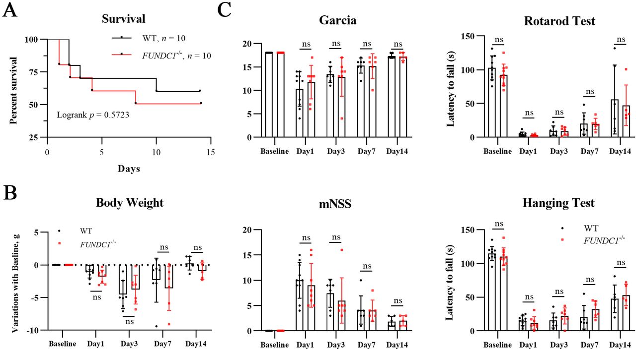
- Figure 2
Absence of FUNDC1 does not influence long-term outcomes of ischaemic stroke. (A) 14-day survival rate after tMCAO. n=10 mice for each genotype. (B–C) Body weight recovery (B) and behavioural tests (C) including Garcia score, mNSS score, rotarod test, hanging test were assessed before the surgery (WT, n=10; FUNDC1−/−, n=10) and at 1 day (WT, n=10; FUNDC1−/−, n=8), 3-day (WT, n=7; FUNDC1−/−, n=7), 7-day (WT, n=6; FUNDC1−/−, n=6), 14-day (WT, n=5; FUNDC1−/−, n=5) after tMCAO. FUNDC1, FUN14 domain-containing 1; mNSS, Modified Neurological Severity Score; tMCAO, transient middle cerebral artery occlusion; WT, wild-type.
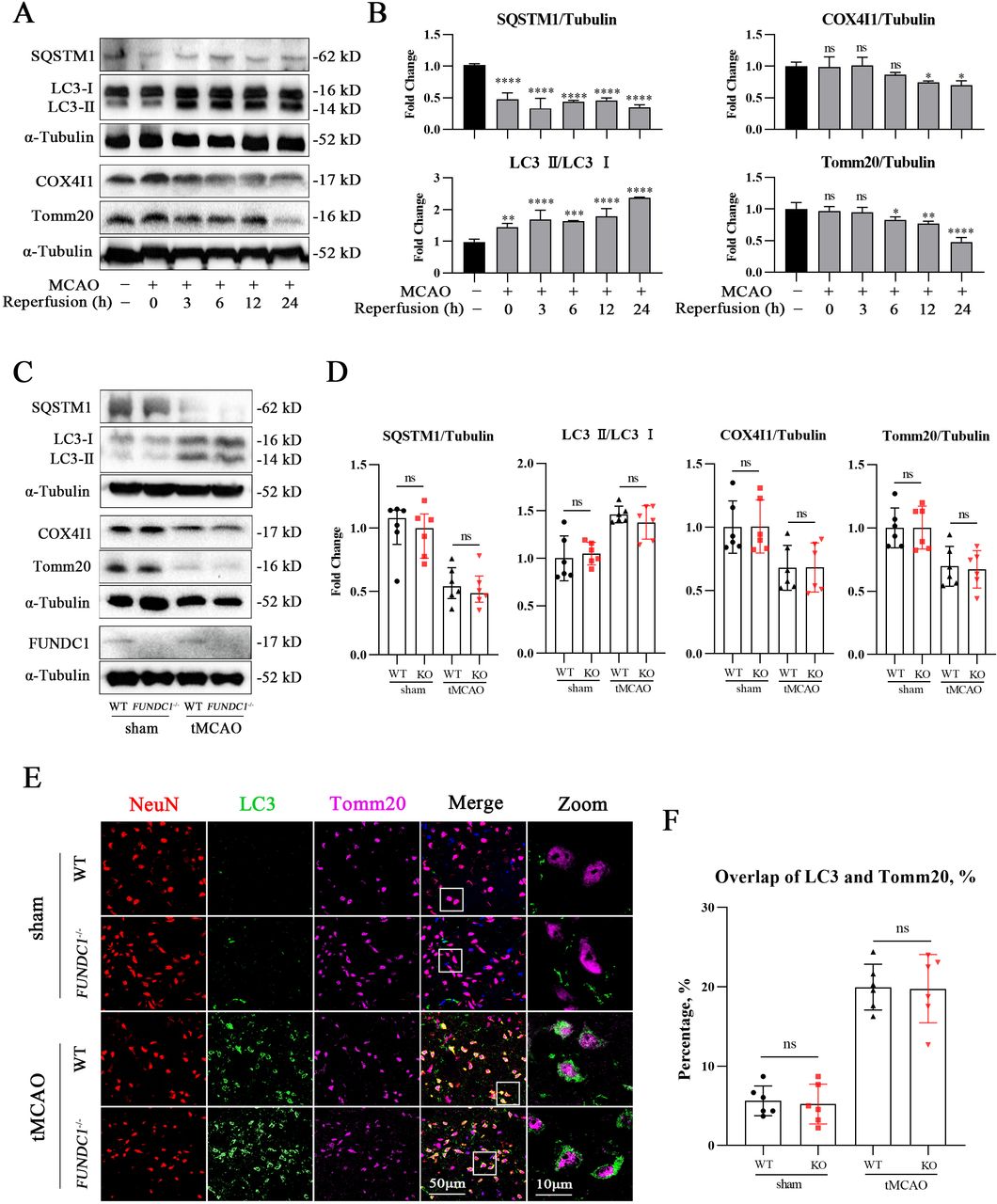
- Figure 3
Deletion of FUNDC1 does not affect neuronal mitophagy in vivo. (A) Time course of mitophagy in vivo was detected by western blotting. (B) Semi-quantification for western blotting detection in panel A. n=3 mice for each time point. (C) WT and FUNDC1−/− mice were subjected to sham surgery or tMCAO for 24 hours. Mitophagy was detected by western blotting. (D) Semi-quantification for western blotting detection in panel C. n=6 mice per group. (E) Brain sections from both genotypes of normal mice or mice with stroke were labelled with NeuN (red), LC3 (green), and Tomm20 (magenta). (F) Quantification of the overlap coefficient of LC3 and Tomm20. n=6 mice per group. *p<0.05, **p<0.01, ***p<0.001, ****p<0.0001. FUNDC1, FUN14 domain-containing 1; KO: Knockout; LC3, light chain 3; SQSTM1: Sequestosome 1; tMCAO, transient middle cerebral artery occlusion; WT, wild-type.
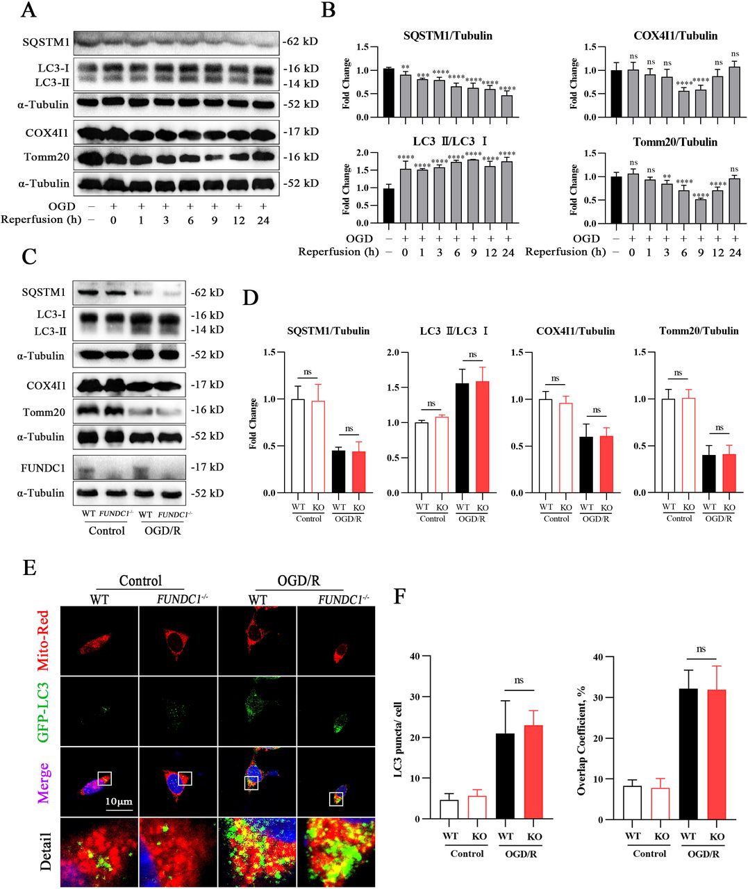
- Figure 4
Loss of FUNDC1 does not affect neuronal mitophagy and mitochondrial qualities in vitro. (A) Time course of neuronal mitophagy in vitro was detected by western blotting. (B) Semi-quantification for western blotting detection in panel A. n=3 for independent experiments. (C) Cortical neurons isolated from WT and FUNDC1–/– mice were subjected to control or OGD/R treatment for 9 hours. Mitophagy was detected by western blotting. (D) Semi-quantification for western blotting detection in panel C. n=3 per independent experiment. (E) Normal-cultured or OGD/R-treated neurons with two genotypes were labelled with MitoTracker (red) and GFP-LC3 (green), then visualised by confocal microscopy. (F) Quantification of the number of LC3 puncta per cell (left) and colocalisation coefficient of mitochondria and LC3 puncta (right). n=3 for independent experiment. **p<0.01, ***p<0.001, ****p<0.0001. FUNDC1, FUN14 domain-containing 1; GFP: Green Fluorescent Protein; KO: Knockout; LC3, light chain 3; OGD/R, oxygen glucose deprivation/reperfusion; SQSTM1: Sequestosome 1; WT, wild-type.
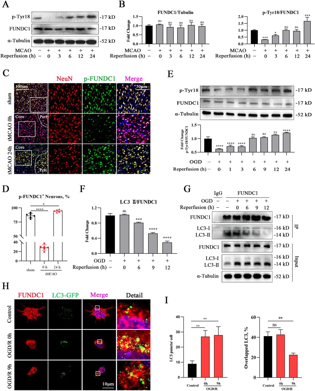
- Figure 5
FUNDC1 is inactivated in later stages of neuronal I/R injury. (A) Time course of FUNDC1 phosphorylation was detected by western blotting in vivo. (B) Semi-quantification for p-Tyr18 and total FUNDC1 in panel A. n=3 mice per time point. (C) Immunostaining of p-FUNDC1 (green) in neurons (red). (D) Quantification for proportion of p-FUNDC1 positive neurons. n=6 mice per time point. (E) Time course of FUNDC1 phosphorylation was detected by western blotting in vitro. n=3 for independent experiments. (F) Semi-quantification for LC3 II interacted with FUNDC1 as is shown in panel G. n=3 for independent experiments. (G) Isolated cortical neurons were subjected to 2 hours of OGD, followed by 0 hours, 6 hours, 9 hours or 12 hours of reperfusion. Interactions between FUNDC1 and LC3 were detected by co-immunoprecipitation. (H) Fluorescent staining of FUNDC1 (red) and GFP-LC3 (green) in isolated neurons subjected to control treatment, OGD/R 0 hours, or OGD/R 9 hours. (I) Quantification of the number of LC3 puncta per cell (left) and percentage of LC3 puncta colocalised with FUNDC1 (right). N=3 for independent experiment. *p<0.05, **p<0.01, ***p<0.001, ****p<0.0001. FUNDC1, FUN14 domain-containing 1; Green Fluorescent Protein; I/R, ischaemia/reperfusion; LC3, light chain 3; OGD/R, oxygen glucose deprivation/reperfusion; tMCAO, transient middle cerebral artery occlusion; WT, wild-type.
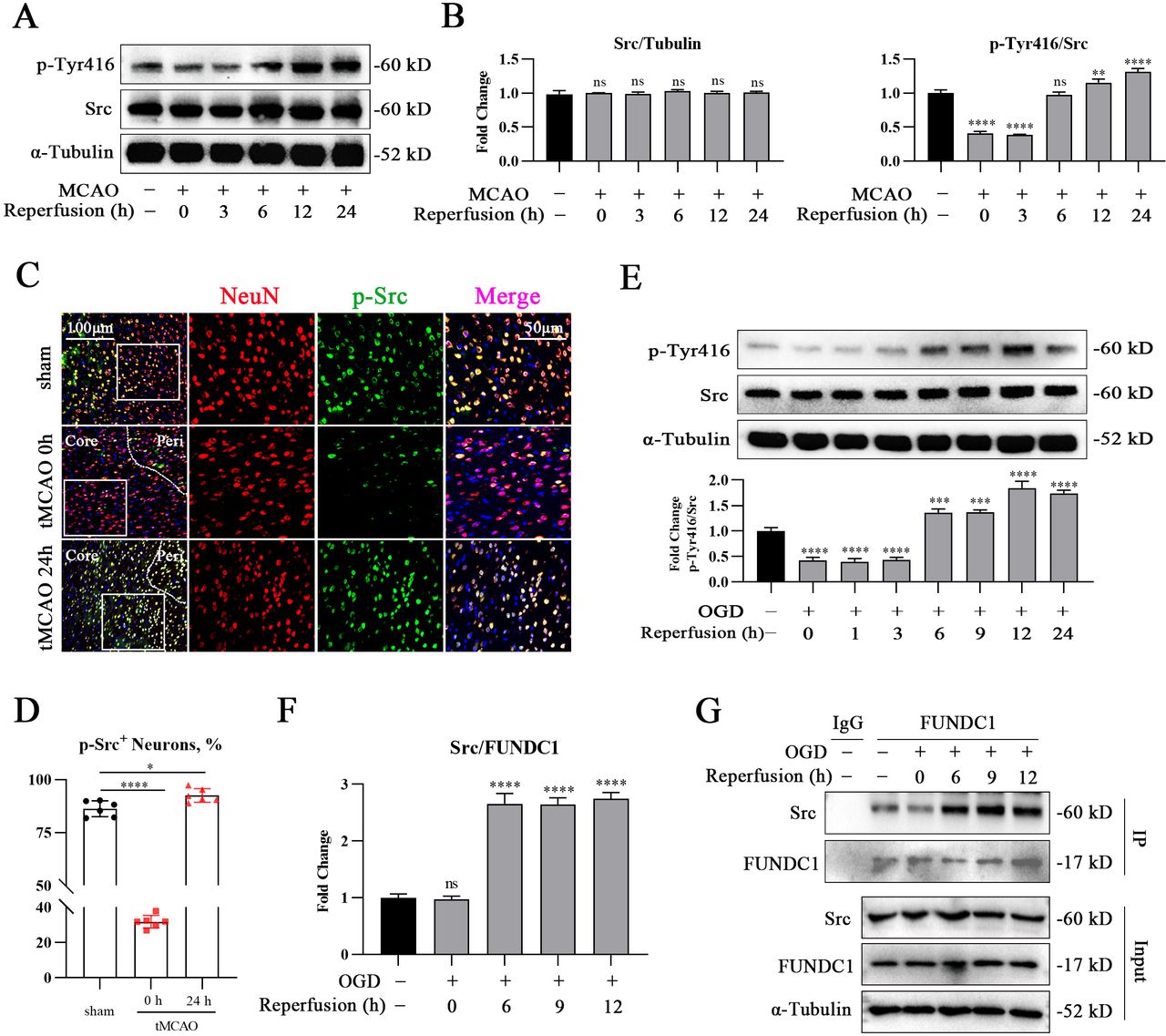
- Figure 6
Src is activated in later stages of neuronal ischaemia/reperfusion injury. (A) Src phosphorylation in vivo at different time points was detected by western blotting. (B) Semi-quantification for p-Tyr416 and total Src in panel A. n=3 mice per time point. (C) Immunostaining of p-Src (green) in neurons (red). (D) Quantification for proportion of p-Src positive neurons. n=6 mice per time point. (E) Src phosphorylation in vitro at different time points was detected by western blotting. n=3 for independent experiments. (F) Semi-quantification for Src interacted with FUNDC1 as is shown in panel G. n=3 for independent experiments. (G) Time course of FUNDC1-Src interaction in isolated neurons were detected by co-immunoprecipitation. *p<0.05, **p<0.01, ***p<0.001, ****p<0.0001. FUNDC1, FUN14 domain-containing 1; OGD, oxygen glucose deprivation; tMCAO, transient middle cerebral artery occlusion.
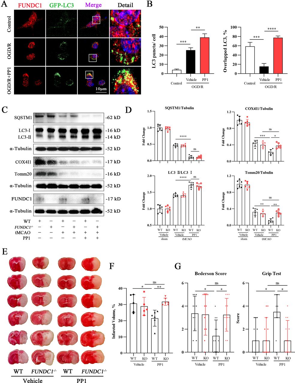
- Figure 7
Pharmacological inhibition of Src rescues FUNDC1-mediated mitophagy in neurons subjected to ischaemia/reperfusion injury. (A) Neurons subjected to OGD/R were treated with vehicle or PP1, and were labelled using antibodies against FUNDC1 (red) and GFP-LC3 (green). (B) Quantification of the number of LC3 puncta per cell (left) and percentage of LC3 puncta colocalised with FUNDC1 (right). n=3 for independent experiment. (C) Mitophagy in mice with both genotypes treated by PP1 in brain samples was detected by western blotting. (D) Semi-quantification for western blotting detection in panel C. n=6 mice per group. (E) Representative TTC staining of brains from vehicle-treated WT mice (n=5), FUNDC1–/– mice (n=5), PP1-treated WT mice (n=6), and PP1-treated FUNDC1–/– mice (n=6). (F) Quantification of infarcted volume. (G) Comparison of neurological deficits among vehicle-treated WT mice (n=11), FUNDC1–/– mice (n=11) PP1-treated WT mice (n=12), and PP1-treated FUNDC1–/– mice (n=12). *p<0.05, **p<0.01, ***p<0.001, ****p<0.0001. FUNDC1, FUN14 domain-containing 1; GFP: Green Fluorescent Protein; OGD/R, oxygen glucose deprivation/reperfusion; KO: Knockout; LC3, light chain 3; SQSTM1: Sequestosome 1; tMCAO, transient middle cerebral artery occlusion; WT, wild-type.
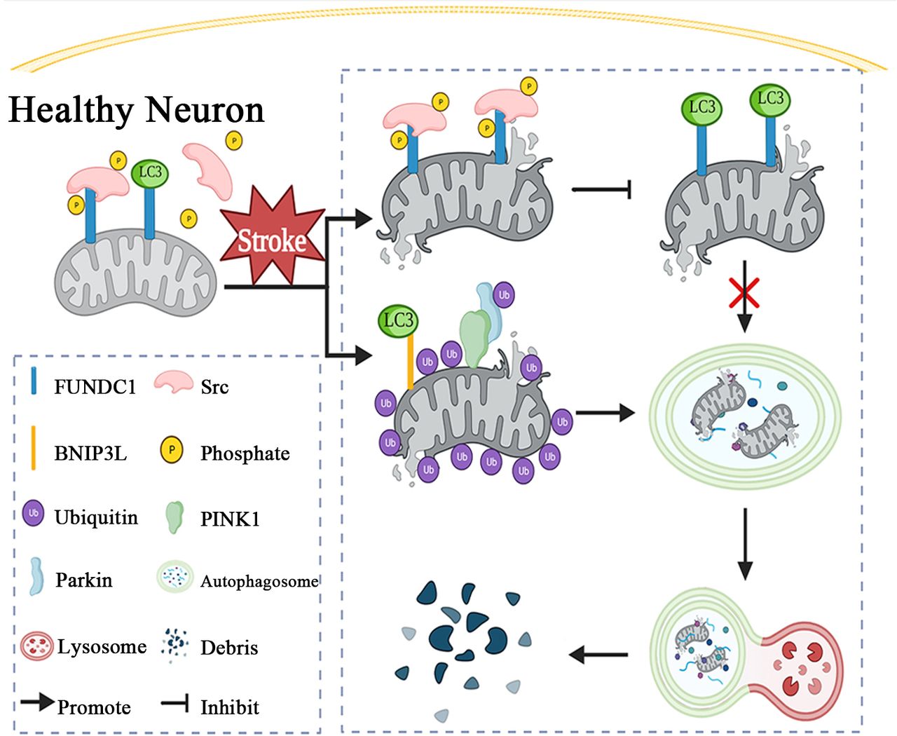
- Figure 8
Working model of FUNDC1 inactivation by Src during neuronal I/R injury (created with BioRender.com). Although neuronal I/R induces mitophagy, hyperactivated Src inactivates FUNDC1, leading to FUNDC1 exclusion from mitophagy. Even in the absence of FUNDC1, neurons can initiate mitophagy through the BNIP3L/Nix and PINK1/Parkin pathways. FUNDC1, FUN14 domain-containing 1; I/R, ischaemia/reperfusion.


Supplementary Materials
Supplementary data
Additional Files
Supplementary Data
This web only file has been produced by the BMJ Publishing Group from an electronic file supplied by the author(s) and has not been edited for content.


