
Main menu

  • Online first
    • Online first
  • Current issue
    • Current issue
  • Archive
    • Archive
  • Submit a paper
    • Online submission site
    • Instructions for authors
  • About the journal
    • About the journal
    • Editorial board
    • Instructions for authors
    • FAQs
    • Chinese Stroke Association
  • Help
    • Contact us
    • Feedback form
    • Reprints
    • Permissions
    • Advertising
  • BMJ Journals

User menu

  • Login

Search

  • Advanced search
  • BMJ Journals
  • Login
  • Facebook
  • Twitter
Stroke and Vascular Neurology

Advanced Search

  • Online first
    • Online first
  • Current issue
    • Current issue
  • Archive
    • Archive
  • Submit a paper
    • Online submission site
    • Instructions for authors
  • About the journal
    • About the journal
    • Editorial board
    • Instructions for authors
    • FAQs
    • Chinese Stroke Association
  • Help
    • Contact us
    • Feedback form
    • Reprints
    • Permissions
    • Advertising
Open Access

MALAT1 promotes platelet activity and thrombus formation through PI3k/Akt/GSK-3β signalling pathway

Yeying Sun, Tao Wang, Yan Lv, Jiahua Li, Xiaoli Jiang, Jing Jiang, Daolai Zhang, Weihua Bian, Chunxiang Zhang
DOI: 10.1136/svn-2022-001498 Published 14 October 2022
Yeying Sun
1College of Pharmacy, Binzhou Medical University, Yantai, Shandong, China
  • Find this author on Google Scholar
  • Find this author on PubMed
  • Search for this author on this site
  • ORCID record for Yeying Sun
Tao Wang
1College of Pharmacy, Binzhou Medical University, Yantai, Shandong, China
  • Find this author on Google Scholar
  • Find this author on PubMed
  • Search for this author on this site
Yan Lv
2College of Life Sciences, Yantai University, Yantai, Shandong, China
  • Find this author on Google Scholar
  • Find this author on PubMed
  • Search for this author on this site
Jiahua Li
1College of Pharmacy, Binzhou Medical University, Yantai, Shandong, China
  • Find this author on Google Scholar
  • Find this author on PubMed
  • Search for this author on this site
Xiaoli Jiang
1College of Pharmacy, Binzhou Medical University, Yantai, Shandong, China
  • Find this author on Google Scholar
  • Find this author on PubMed
  • Search for this author on this site
Jing Jiang
1College of Pharmacy, Binzhou Medical University, Yantai, Shandong, China
  • Find this author on Google Scholar
  • Find this author on PubMed
  • Search for this author on this site
Daolai Zhang
1College of Pharmacy, Binzhou Medical University, Yantai, Shandong, China
  • Find this author on Google Scholar
  • Find this author on PubMed
  • Search for this author on this site
Weihua Bian
1College of Pharmacy, Binzhou Medical University, Yantai, Shandong, China
  • Find this author on Google Scholar
  • Find this author on PubMed
  • Search for this author on this site
  • ORCID record for Weihua Bian
Chunxiang Zhang
1College of Pharmacy, Binzhou Medical University, Yantai, Shandong, China
3Department of Cardiology, Southwest Medical University, Luzhou, Sichuan, China
  • Find this author on Google Scholar
  • Find this author on PubMed
  • Search for this author on this site
  • Article
  • Figures & Data
  • eLetters
  • Info & Metrics
  • PDF
Loading

Article Figures & Data

Figures

  • Supplementary Materials
  • Additional Files
  • Figure 1
    • Download figure
    • Open in new tab
    • Download powerpoint
    Figure 1

    MALAT1 is present in the cytoplasm of MEG01 and downregulated in activated MEG01 cells. (A) RNA fluorescence in situ hybridisation (FISH) of the MALAT1 and 18S in MEG01 cells. Scale bar, 20 µm. There were three separate tests carried out, and each image represents a different one of those experiments. (B) MALAT1 distribution in the nucleus and cytoplasm of MEG01 cells by RT-PCR. Y-axis represents the mean of CT value. GAPDH is used as a cytoplasmic reference. (C) After treating MEG-01 cells with thrombin at a concentration of 1 U/mL for 6, 12, and 24 hours, relative MALAT1 expression was evaluated (**p<0.01) in these cells on collagen-coated coverslips. RT-PCR, reverse transcription PCR.
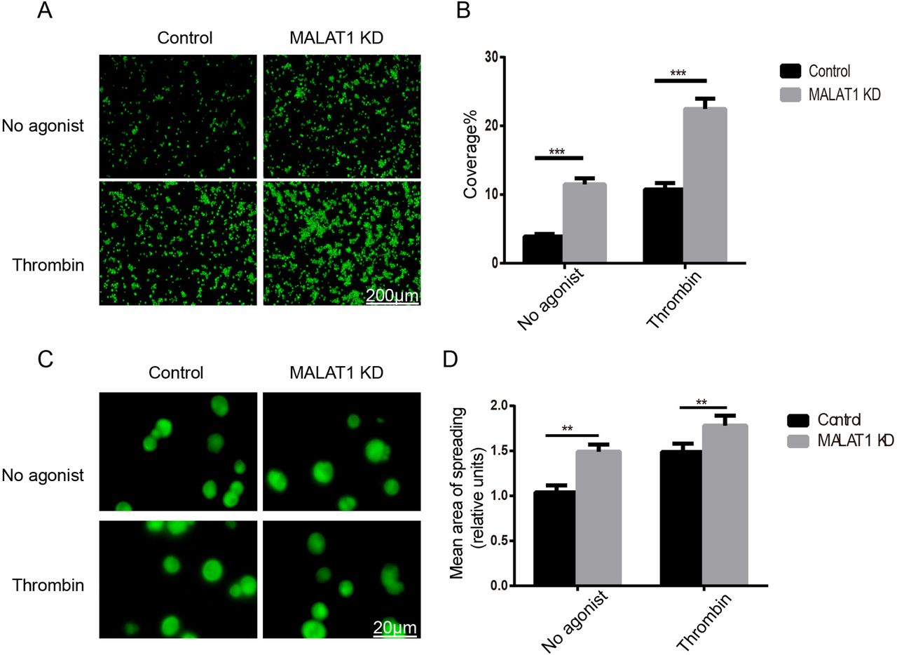
  • Figure 2
    • Download figure
    • Open in new tab
    • Download powerpoint
    Figure 2

    MALAT1 knockdown (KD) in CD34+ megakaryocytes represses adhesion and spreading. Each experiment was performed in triplicate. (A) Comparison of the capacity of MALAT1 KD and control CD34+megakaryocytes (green) to adhere to collagen-coated coverslips following stimulation with or without thrombin 1 U/mL (scale bar, 200 µm). (B) Statistical analysis of the adhesion area of CD34+ megakaryocytes in collagen-coated coverslips. Values are mean±SD (***p<0.001). (C) Comparison of CD34+ megakaryocyte spreading potential on collagen-coated coverslips between MALAT1 KD and control groups without and with 1 U/mL thrombin (scale bar, 20 m). (D) Spreading area was compared with that of platelets of the wild type (**p<0.01), and the mean±SEM was plotted. Around 30 platelets from each genotype were counted on average.
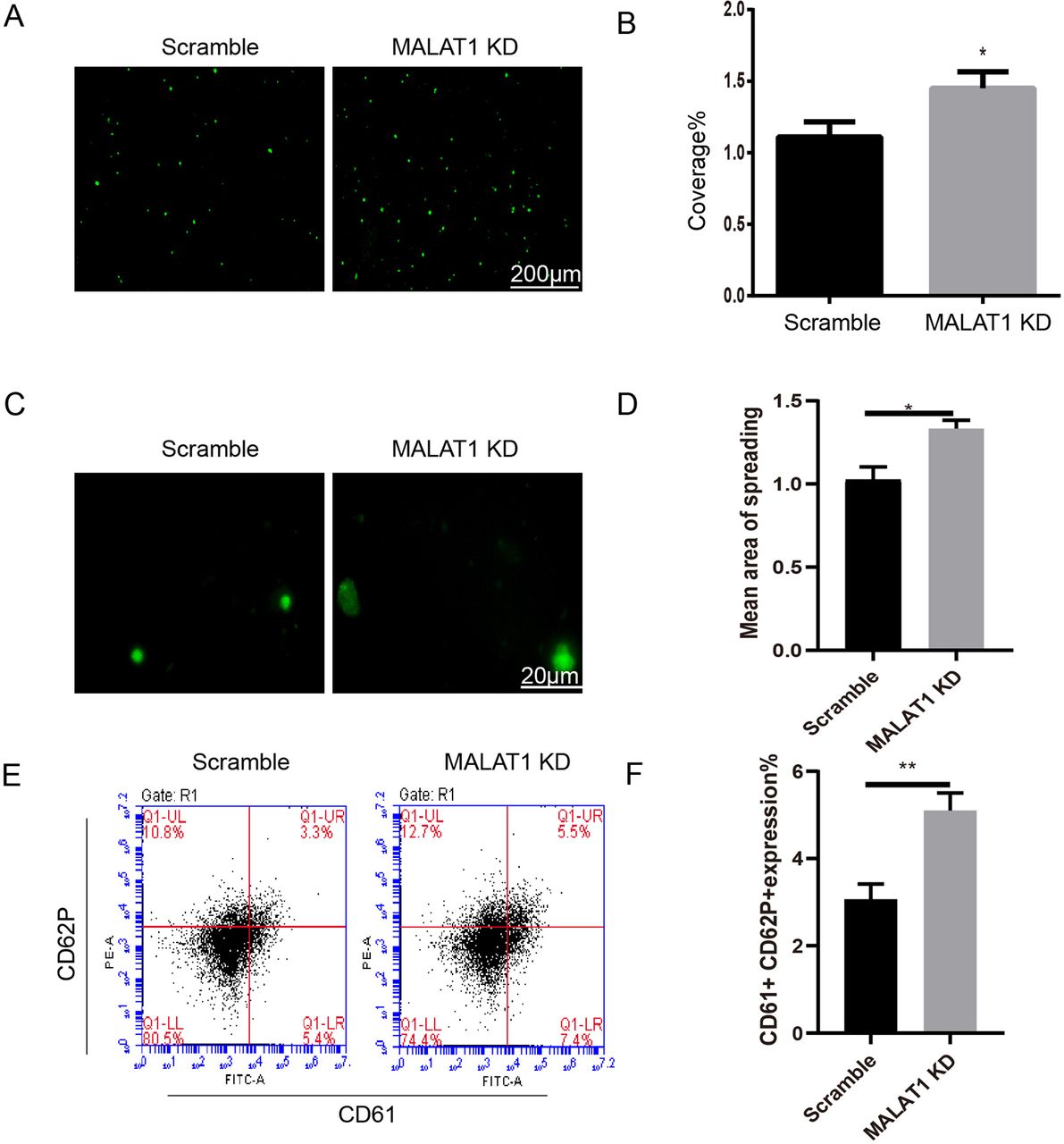
  • Figure 3
    • Download figure
    • Open in new tab
    • Download powerpoint
    Figure 3

    MALAT1 represses PLP adhesion, spreading and activity. (A) Adhesion of PLPs (green) from MALAT1 KD and control CD34+ megakaryocytes after treatment by thrombin 1 U/mL (scale bar, 200 μm). Three separate sets of data were collected for each experiment. (B) A statistical examination of the adhesion area of PLPs in coverslips that were coated with collagen. The values are shown as the mean±SD (*p<0.05). (C) A comparison of the capacity of PLPs to spread derived from MALAT1 kD CD34+ megakaryocytes and the scramble in the presence of thrombin at a concentration of 1 U/mL on coverslips coated with collagen (scale bar, 20 µm). (D) Histogram showing the relative area of PLPs for MALAT1 KD and scramble. Data are from three independent experiments. Error bars denote SD *p<0.05. (E) Flow cytometric analysis of CD61 and CD62P expressions on the PLPs isolated from MALAT1 kD megakaryocytes and scramble. (F) Histogram showing the proportions of CD61+CD62P+ cells for MALAT1 kD megakaryocytes and scramble group. Data are from three independent experiments. Error bars denote SD **p<0.01. KD, knockdown; PLPs, platelet-like particles.
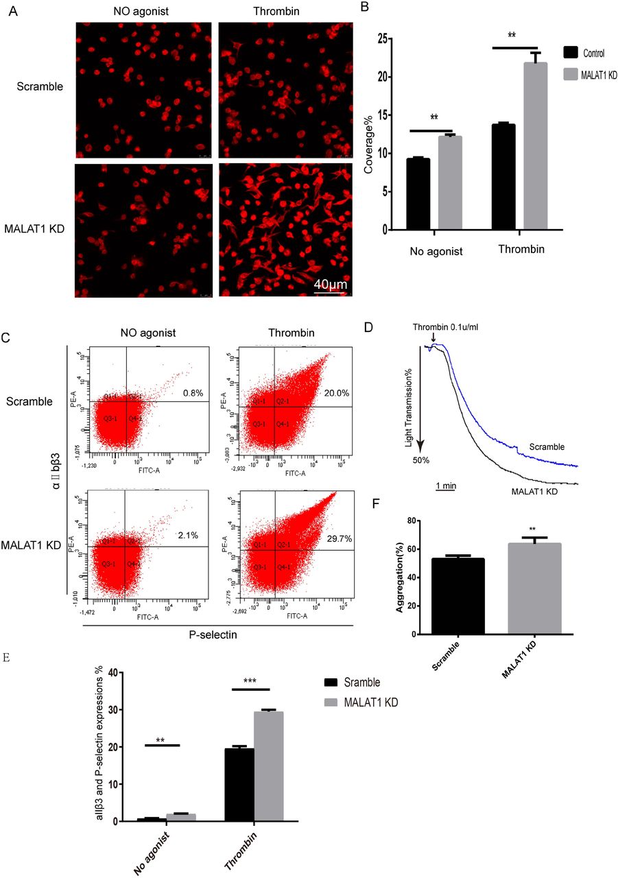
  • Figure 4
    • Download figure
    • Open in new tab
    • Download powerpoint
    Figure 4

    MALAT1 regulates platelet spreading, aggregation, and activity. (A) Platelets with a deficiency in MALAT1 and platelets with a wild-type genotype (WT) were spread out over fibrinogen-coated coverslips with or without thrombin (0.1 U/mL). (B) Quantification analysis of the spreading area of MALAT1-deficient and WT platelets. Values are mean±SD (**p<0.001). (C) Flow cytometric analysis of integrin aIIβ3 and P-selectin expressions of MALAT1-deficient and WT platelets in the absence or presence of thrombin (0.1 U/mL), using JON/A PE-labelled JON/A antibody and FITC-labelled Wug.E9 antibody (n=6). (D) Aggregation of washed MALAT1-deficient and WT platelets using thrombin (0.1 U/mL). (E) Histogram showing the proportions of aIIβ3 and P-selectin cells for MALAT1 KD megakaryocytes and scramble group. Data are from three independent experiments. Error bars denote SD **p<0.01, ***p<0.001. (F) Panel D depicts the percentage of platelet maximum aggregation based on the findings of at least five separate studies, and the data are presented as the mean±SD (**p<0.01). FITC, fluorescein isothiocyanate; KD, knockdown.
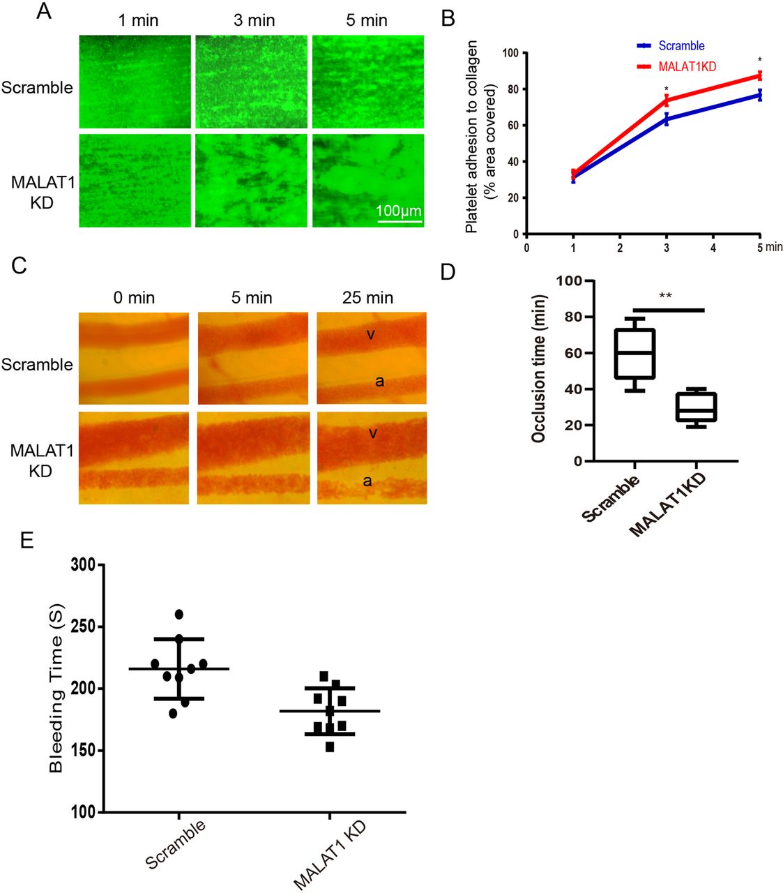
  • Figure 5
    • Download figure
    • Open in new tab
    • Download powerpoint
    Figure 5

    Downregulated MALAT1 in platelets increases thrombus formation in vitro and in vivo. (A) Photomicrographs that show the progression of platelet adhesion from MALAT1 KD and scramble mice on collagen. Blood from MALAT1 KD and scrambling animals was collected in sodium citrate (3.8% w/v) and fluorescently tagged with calcein (1 µM) for 1 hour before being perfused through fibrillar collagen-coated bioflux plates at a shear rate of 40 dynes/cm2 for 5 min. (B) Dot plot comparing the area coverage of platelets derived from scramble mice and MALAT1 knockout mice. There were a total of three separate trials carried out. *p<0.05 (C) Images showing representative stages of thrombus formation at 0 min, 5 min, and 25 min after an injury caused by FeCl3 to mesenteric arterioles in MALAT1 KD mice and scrambling mice. A, arteriole; V, venule. (D) The occlusion periods for arteriole thrombosis produced by FeCl3 in scramble and MALAT1 KD mice are displayed as box plots (n=5). **p<0.01. (E) MALAT1 KD accelerates haemostatic plug formation in mice. horizontal lines represent the mean (eight individuals in each group). KD, knockdown.
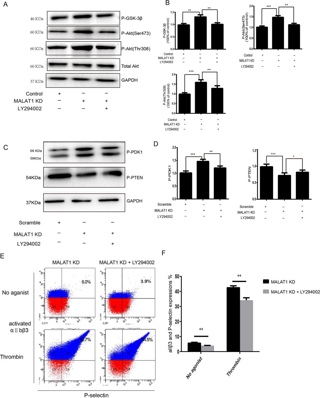
  • Figure 6
    • Download figure
    • Open in new tab
    • Download powerpoint
    Figure 6

    MALAT1 deficiency in platelets activates PI3k/Akt signal pathway. (A) Western blot analysis of WT and MALAT1-deficient platelets in the absence or presence of LY294002 (50 nmol/kg) with antibodies that recognise phosphorylated GSK-3β, phosphorylated Akt Thr308, phosphorylated Akt Ser473, and total Akt. (B) Western blot results were quantified and presented as mean±SD (**p<0.01, ***p<0.001). (C) Analysis of phosphorylated PDK1 and PTEN by Western blotting in MALAT1-deficient and WT platelets in the absence or presence of LY294002 (50 nmol/kg), respectively. (D) The findings of the Western blot were analysed quantitatively and displayed as the mean±SD (*p<0.05, **p<0.01, ***p<0.001). (E) Flow cytometric examination of integrin aIIβ3 and P-selectin expressions with JON/A PE-labelled JON/A antibody and FITC-labelled Wug.E9 antibody (n=6). This was done in the absence or presence of the PI3K inhibitor LY294002. (F) A histogram depicting the percentages of cells expressing aIIβ3 and P-selectin with or without the PI3K inhibitor LY294002. Data are from three independent experiments. Error bars denote SD **p<0 .01. WT, wild-type.
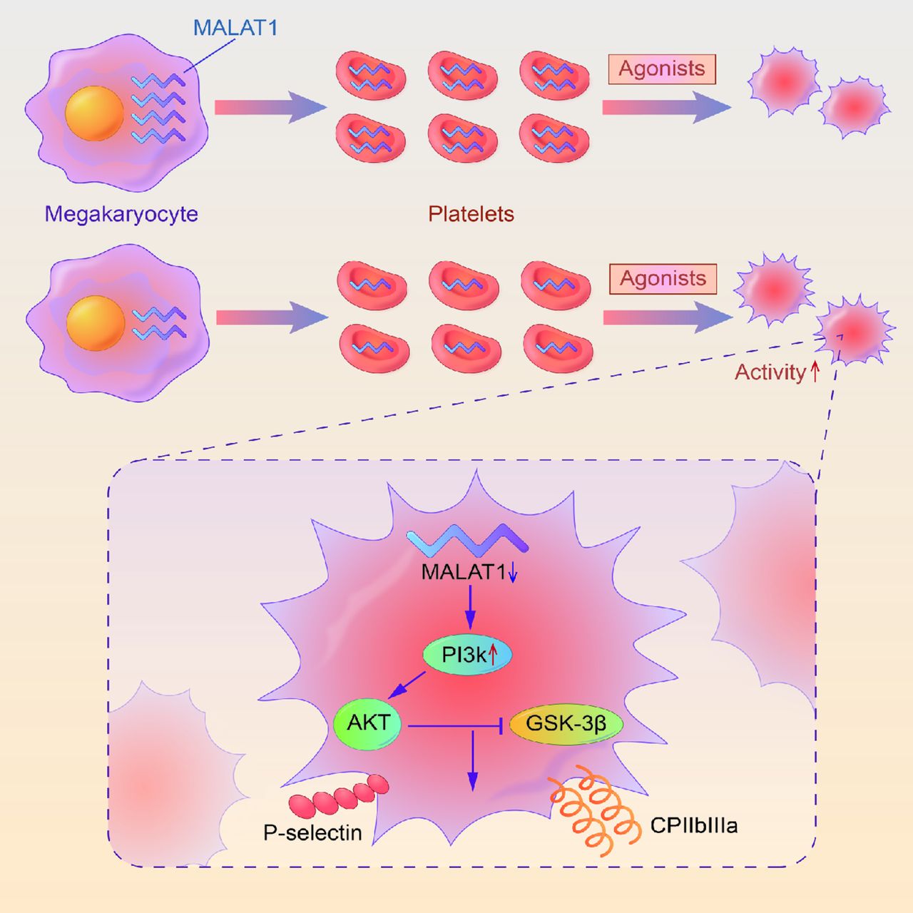
  • Figure 7
    • Download figure
    • Open in new tab
    • Download powerpoint
    Figure 7

    MALAT1 regulates platelet activity through PI3k/Akt/GSK-3β pathway. MALAT1 is a potent negative regulator of platelet activity. In the absence of this lncRNA, integrin outside-in signalling can be activated by PI3k/Akt/GSK-3β pathway.

Supplementary Materials

  • Figures
  • Additional Files
  • Supplementary data

    [svn-2022-001498supp001.pdf]

Additional Files

  • Figures
  • Supplementary Materials
  • Supplementary Data

    This web only file has been produced by the BMJ Publishing Group from an electronic file supplied by the author(s) and has not been edited for content.

    • Data supplement 1
PreviousNext
Back to top
Vol 10 Issue 1 Table of Contents
Stroke and Vascular Neurology: 10 (1)
  • Table of Contents
  • Table of Contents (PDF)
  • About the Cover
  • Index by author
  • Ed Board (PDF)
Email

Thank you for your interest in spreading the word on Stroke and Vascular Neurology.

NOTE: We only request your email address so that the person you are recommending the page to knows that you wanted them to see it, and that it is not junk mail. We do not capture any email address.

Enter multiple addresses on separate lines or separate them with commas.
MALAT1 promotes platelet activity and thrombus formation through PI3k/Akt/GSK-3β signalling pathway
(Your Name) has sent you a message from Stroke and Vascular Neurology
(Your Name) thought you would like to see the Stroke and Vascular Neurology web site.
CAPTCHA
This question is for testing whether or not you are a human visitor and to prevent automated spam submissions.
Print
Alerts
Sign In to Email Alerts with your Email Address
Citation Tools
MALAT1 promotes platelet activity and thrombus formation through PI3k/Akt/GSK-3β signalling pathway
Yeying Sun, Tao Wang, Yan Lv, Jiahua Li, Xiaoli Jiang, Jing Jiang, Daolai Zhang, Weihua Bian, Chunxiang Zhang
Stroke and Vascular Neurology Oct 2022, svn-2022-001498; DOI: 10.1136/svn-2022-001498

Citation Manager Formats

  • BibTeX
  • Bookends
  • EasyBib
  • EndNote (tagged)
  • EndNote 8 (xml)
  • Medlars
  • Mendeley
  • Papers
  • RefWorks Tagged
  • Ref Manager
  • RIS
  • Zotero
Cite This
  • APA
  • Chicago
  • Endnote
  • MLA
Loading
MALAT1 promotes platelet activity and thrombus formation through PI3k/Akt/GSK-3β signalling pathway
Yeying Sun, Tao Wang, Yan Lv, Jiahua Li, Xiaoli Jiang, Jing Jiang, Daolai Zhang, Weihua Bian, Chunxiang Zhang
Stroke and Vascular Neurology Oct 2022, svn-2022-001498; DOI: 10.1136/svn-2022-001498
Download PDF

Share
MALAT1 promotes platelet activity and thrombus formation through PI3k/Akt/GSK-3β signalling pathway
Yeying Sun, Tao Wang, Yan Lv, Jiahua Li, Xiaoli Jiang, Jing Jiang, Daolai Zhang, Weihua Bian, Chunxiang Zhang
Stroke and Vascular Neurology Oct 2022, svn-2022-001498; DOI: 10.1136/svn-2022-001498
Reddit logo Twitter logo Facebook logo Mendeley logo
  • Tweet Widget
  • Facebook Like
  • Google Plus One
  • Article
    • Abstract
    • Introduction
    • Materials and methods
    • Results
    • Discussion
    • Data availability statement
    • Ethics statements
    • Footnotes
    • References
  • Figures & Data
  • eLetters
  • Info & Metrics
  • PDF

Related Articles

Cited By...

More in this TOC Section

  • Learning curve and embolisation strategy in single-stage surgery combined embolisation and microsurgery for brain arteriovenous malformations: results from a nationwide multicentre prospective registry study
  • Stepwise improvement in intracerebral haematoma expansion prediction with advanced imaging: a comprehensive comparison of existing scores
  • Thrombus iodine-based perviousness is associated with recanalisation and functional outcomes in endovascular thrombectomy
Show more Original research

Similar Articles


CONTENT

  • Latest content
  • Current issue
  • Archive
  • eLetters
  • Sign up for email alerts
  • RSS

JOURNAL

  • About the journal
  • Editorial board
  • Recommend to librarian
  • Chinese Stroke Association

AUTHORS

  • Instructions for authors
  • Submit a paper
  • Track your article
  • Open Access at BMJ

HELP

  • Contact us
  • Reprints
  • Permissions
  • Advertising
  • Feedback form

© 2025 Chinese Stroke Association