
Main menu

  • Online first
    • Online first
  • Current issue
    • Current issue
  • Archive
    • Archive
  • Submit a paper
    • Online submission site
    • Instructions for authors
  • About the journal
    • About the journal
    • Editorial board
    • Instructions for authors
    • FAQs
    • Chinese Stroke Association
  • Help
    • Contact us
    • Feedback form
    • Reprints
    • Permissions
    • Advertising
  • BMJ Journals

User menu

  • Login

Search

  • Advanced search
  • BMJ Journals
  • Login
  • Facebook
  • Twitter
Stroke and Vascular Neurology

Advanced Search

  • Online first
    • Online first
  • Current issue
    • Current issue
  • Archive
    • Archive
  • Submit a paper
    • Online submission site
    • Instructions for authors
  • About the journal
    • About the journal
    • Editorial board
    • Instructions for authors
    • FAQs
    • Chinese Stroke Association
  • Help
    • Contact us
    • Feedback form
    • Reprints
    • Permissions
    • Advertising
Open Access

M2 middle cerebral artery dissection on 7T MRI

Xuewei Xie, Zhe Zhang, Qingle Kong, Hui Qu, Jing Jing
DOI: 10.1136/svn-2022-001557 Published 14 July 2022
Xuewei Xie
1Department of Neurology, Beijing Tiantan Hospital, Capital Medical University, Beijing, China
2China National Clinical Research Center for Neurological Diseases, Beijing, China
  • Find this author on Google Scholar
  • Find this author on PubMed
  • Search for this author on this site
  • ORCID record for Xuewei Xie
Zhe Zhang
2China National Clinical Research Center for Neurological Diseases, Beijing, China
3Tiantan Neuroimaging Center of Excellence, Beijing, China
  • Find this author on Google Scholar
  • Find this author on PubMed
  • Search for this author on this site
Qingle Kong
4MR Collaboration, Siemens Healthineers Ltd, Beijing, China
  • Find this author on Google Scholar
  • Find this author on PubMed
  • Search for this author on this site
Hui Qu
1Department of Neurology, Beijing Tiantan Hospital, Capital Medical University, Beijing, China
2China National Clinical Research Center for Neurological Diseases, Beijing, China
  • Find this author on Google Scholar
  • Find this author on PubMed
  • Search for this author on this site
Jing Jing
1Department of Neurology, Beijing Tiantan Hospital, Capital Medical University, Beijing, China
2China National Clinical Research Center for Neurological Diseases, Beijing, China
3Tiantan Neuroimaging Center of Excellence, Beijing, China
  • Find this author on Google Scholar
  • Find this author on PubMed
  • Search for this author on this site
  • Article
  • Figures & Data
  • eLetters
  • Info & Metrics
  • PDF
Loading

Article Figures & Data

Figures

  • Figure 1
    • Download figure
    • Open in new tab
    • Download powerpoint
    Figure 1

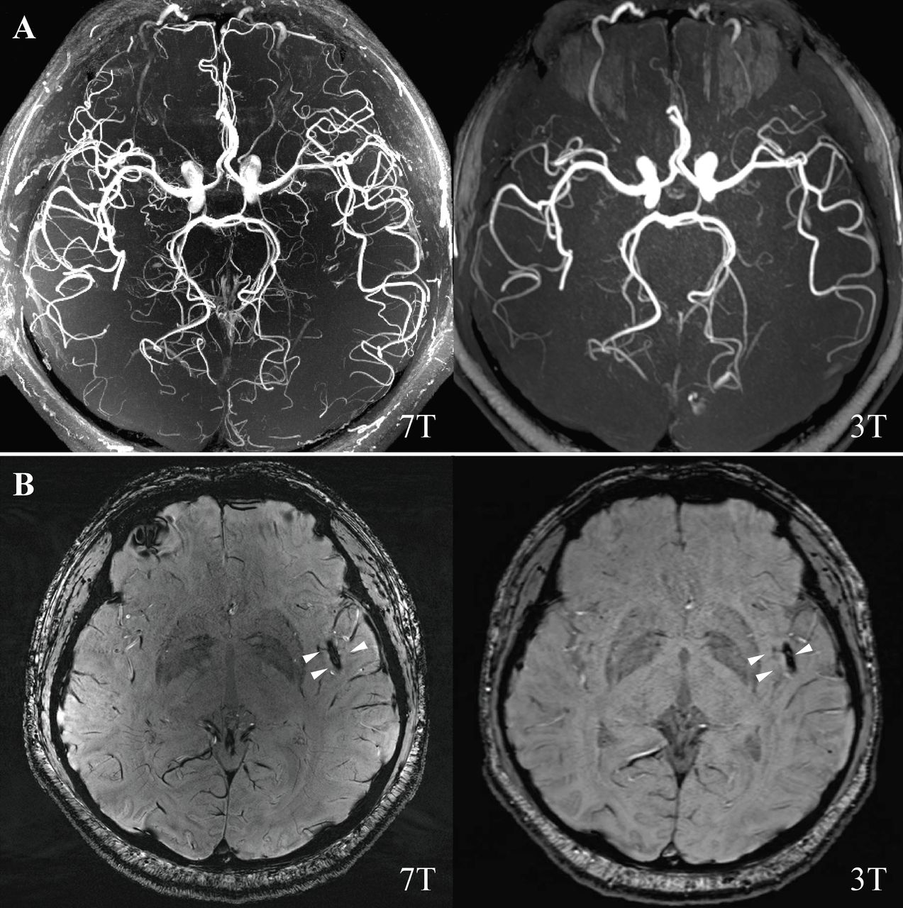
    (A) 7T and 3T magnetic resonance angiography. (B) 7T and 3T SWI were showing blooming hypointense signals (susceptibility vessel sign) in the distal M2 segment of the left MCA (white triangle). MCA, middle cerebral artery; SWI, susceptibility-weighted imaging.

  • Figure 2
    • Download figure
    • Open in new tab
    • Download powerpoint
    Figure 2

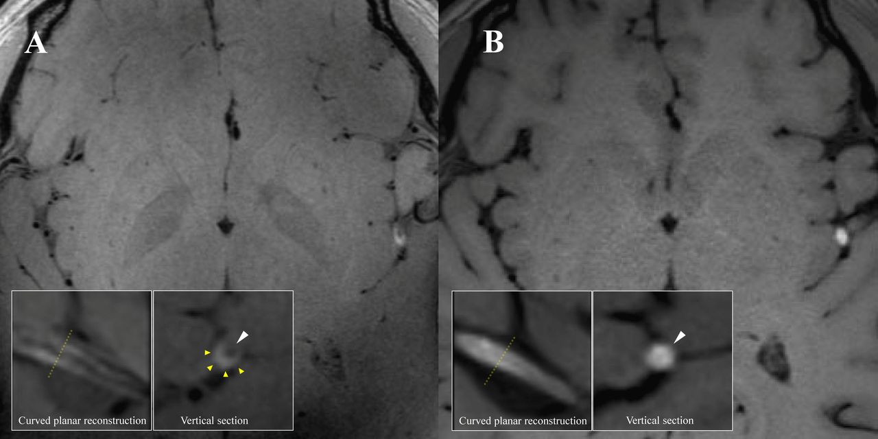
    (A) 7T 3D T1-SPACE MRI (MAGNETOM Terra, Siemens Healthcare, Erlangen, Germany. Voxel size=0.4×0.4×0.4 mm3) showing left distant M2 segment MCA mural haematoma and residual lumen, and axial reformatted images showing a semilunar hyperintense signal caused by a mural haematoma (yellow triangle) and an eccentric hypointense signal attributable to the residual lumen (white triangle). (B) 3T 3D T1-SPACE MRI (MAGNETOM Prisma, Siemens Healthcare, Erlangen, Germany. Voxel size=0.54×0.54×0.54 mm3) showing left distant M2 segment MCA intraluminal thrombus formation and axial reformatted images showing only hyperintense signal (white triangle) with an unclear residual lumen. MCA, middle cerebral artery.

PreviousNext
Back to top
Vol 10 Issue 1 Table of Contents
Stroke and Vascular Neurology: 10 (1)
  • Table of Contents
  • Table of Contents (PDF)
  • About the Cover
  • Index by author
  • Ed Board (PDF)
Email

Thank you for your interest in spreading the word on Stroke and Vascular Neurology.

NOTE: We only request your email address so that the person you are recommending the page to knows that you wanted them to see it, and that it is not junk mail. We do not capture any email address.

Enter multiple addresses on separate lines or separate them with commas.
M2 middle cerebral artery dissection on 7T MRI
(Your Name) has sent you a message from Stroke and Vascular Neurology
(Your Name) thought you would like to see the Stroke and Vascular Neurology web site.
CAPTCHA
This question is for testing whether or not you are a human visitor and to prevent automated spam submissions.
Print
Alerts
Sign In to Email Alerts with your Email Address
Citation Tools
M2 middle cerebral artery dissection on 7T MRI
Xuewei Xie, Zhe Zhang, Qingle Kong, Hui Qu, Jing Jing
Stroke and Vascular Neurology Jul 2022, svn-2022-001557; DOI: 10.1136/svn-2022-001557

Citation Manager Formats

  • BibTeX
  • Bookends
  • EasyBib
  • EndNote (tagged)
  • EndNote 8 (xml)
  • Medlars
  • Mendeley
  • Papers
  • RefWorks Tagged
  • Ref Manager
  • RIS
  • Zotero
Cite This
  • APA
  • Chicago
  • Endnote
  • MLA
Loading
M2 middle cerebral artery dissection on 7T MRI
Xuewei Xie, Zhe Zhang, Qingle Kong, Hui Qu, Jing Jing
Stroke and Vascular Neurology Jul 2022, svn-2022-001557; DOI: 10.1136/svn-2022-001557
Download PDF

Share
M2 middle cerebral artery dissection on 7T MRI
Xuewei Xie, Zhe Zhang, Qingle Kong, Hui Qu, Jing Jing
Stroke and Vascular Neurology Jul 2022, svn-2022-001557; DOI: 10.1136/svn-2022-001557
Reddit logo Twitter logo Facebook logo Mendeley logo
  • Tweet Widget
  • Facebook Like
  • Google Plus One
  • Article
    • Ethics statements
    • Footnotes
    • References
  • Figures & Data
  • eLetters
  • Info & Metrics
  • PDF

Related Articles

Cited By...

More in this TOC Section

  • Cerebrovascular cinematic rendering of multisystemic smooth muscle dysfunction syndrome
  • Pictures of stroke and cerebral vasculature: novel insights and detailed visualisation of U-shaped elastin distribution and disorganised fibre arrangement at carotid bifurcations by advanced techniques
Show more Pictures of stroke and cerebral vasculature

Similar Articles


CONTENT

  • Latest content
  • Current issue
  • Archive
  • eLetters
  • Sign up for email alerts
  • RSS

JOURNAL

  • About the journal
  • Editorial board
  • Recommend to librarian
  • Chinese Stroke Association

AUTHORS

  • Instructions for authors
  • Submit a paper
  • Track your article
  • Open Access at BMJ

HELP

  • Contact us
  • Reprints
  • Permissions
  • Advertising
  • Feedback form

© 2025 Chinese Stroke Association