

Article Figures & Data
- Figure 1
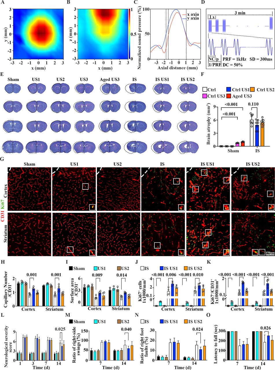
Establishing optimal LIFUS parameters for neuronal repair and remodelling in MCAO mice. (A) 2D distribution of the ultrasound field in oblique and longitudinal cross-sections. The x-axis and y-axis of the ultrasound spot were ~4.5 mm and ~5 mm, respectively. (B) Depth of ultrasound irradiation was 4 mm. (C) X-Y diameter was 2 mm when normalised sound pressure was more than 50% of the total irradiation energy. (D) Schematic diagram of ultrasound, PRF was 1 kHz and 1/PRF was 1 s, SD was 300 ms, and DC was 50%. (E) Cresyl violet-stained brain sections and (F) quantification of atrophy volume at 14 days following MCAO. Dashed lines indicated brain atrophy area, (n=3–7 mice/group). (G) Representative CD31 (red) and Ki67 (green) immunostaining images in the perifocal region. (H) Quantitative analysis of capillary number, (I) surface area, (J) Ki67+, and (K) Ki67+/CD31+ signals in the perifocal region of ipsilateral hemisphere after 14 days following MCAO in mice, (n=4 mice/group. Scale bar=150 µm). (L) mNSS, (M) tail suspension, (N) grid walking and (O) rotarod test for neurobehavioural outcomes in each group (n=3 mice/group in sham groups, n=10–12 mice/group in the IS groups). US1, −2 to –3=mice treated with different dose of ultrasound. IS, ischaemic stroke mice; IS US, ischaemic stroke mice treated with US. Data are mean ± SD. LIFUS, low-intensity focused ultrasound stimulation; mNSS, modified neurological severity score; PRF, pulse repetition frequency; SD, sonication duration.
- Figure 2
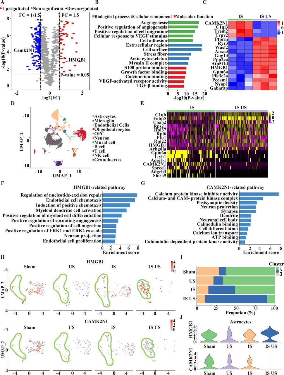
LIFUS upregulated HMGB1 and downregulated CAMK2N1 in a new cluster of astrocytes. (A) Volcano plot demonstrated fold change of protein level of HMGB1and CAMK2N1 in the IS US group compared with the IS group. (B) Bar chart of GO terms showed angiogenesis and synapse related pathways including BP (biological process, green), CC (cell component, blue), and MF (molecular function, red). (C) Heatmap showed different protein expression in the IS US group compared with IS group. (D) UMAP plot showed the expression profiles in the left ipsilateral by clustering cell types in the ipsilateral hemisphere of mouse brain. (E) Heatmap showed differentially expressed genes (DEGs) in the IS US group compared with IS group of astrocytes. (F) Bar chart of GO terms showed enriched HMGB1 and (G) CAMK2N1-related pathways of astrocytes in the IS US group. (H) Expression profiles of HMGB1 and CAMK2N1 in astrocytes organised into groups, and coloured based on gene expression patterns. (I) Bar chart showed the proportions of three subgroups in four different groups. (J) Violin plots represented the expression distributions of HMGB1 and CAMK2N1 in astrocytes organised into groups. GO, gene ontology; IS, ischaemic stroke mice; IS US, ischaemic stroke mice treated with US; US, mice treated with ultrasound; LIFUS, low-intensity focused ultrasound stimulation; UMAP, uniform manifold approximation and projection; VEGF, vascular endothelial growth factor.
- Figure 3
Inhibiting astrocytic HMGB1 attenuated angiogenesis and reversed neurobehavioural outcomes after LIFUS in MCAO mice. (A) Representative in situ hybridisation images of HMGB1 (green) signals and GFAP+ astrocytes (red) in the IS scramble, IS US scramble, IS US sh (HMGB1), and IS US gf-sh (HMGB1) mice. Scale bar=75 µm. (B) Corresponding semiquantification of HMGB1 mRNA expression level in different groups and (C) percentile of astrocytes (n = 3 mice/group). (D) Western blotting (E) and quantification of HMGB1, VEGFA and FGF2, protein levels (from left to right, normalised to corresponding sham) in ipsilateral mice brain, n=4 mice/group for all groups. (F) mNSS, (G) tail suspension, (H) grid walking and (I) rotarod test of neurobehavioural outcomes in each group (n=3 mice/group in sham groups, n=12 mice/group in the IS groups). IS, ischaemic stroke mice; IS US, ischaemic stroke mice treated with US; US, mice treated with ultrasound. Data are mean ± SD. LIFUS, low-intensity focused ultrasound stimulation.
- Figure 4
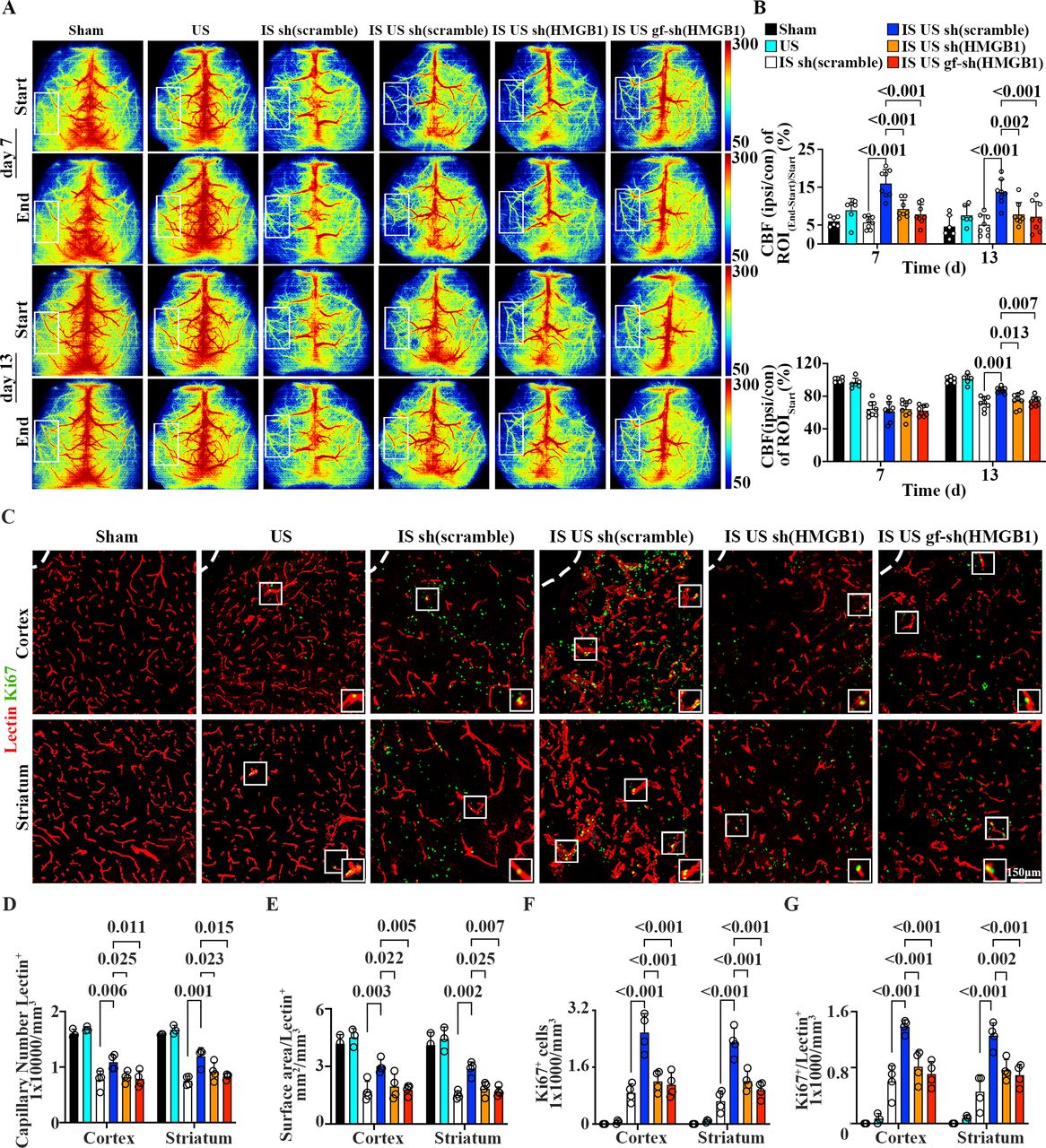
LIFUS upregulated astrocytic HMGB1 via increased CBF and lectin+ microvessels in MCAO mice. (A) Representative images showed immediate CBF changes and endpoint CBF of ROI at 7 days and 13 days by laser speckle imaging in the sham, sham US, IS scramble, IS US scramble, IS US sh (HMGB1), IS US gf-sh (HMGB1) groups. (B) Quantification of CBF normalised to sham. Start row was the quantification of CBF at 7 days and 13 days, respectively. End row was the quantification of immediate CBF changes followed ultrasound at 7 days and 13 days respectively, (n=6 mice/group in the sham groups, n=10 mice/group in the IS groups). (C) Representative lectin (red) and Ki67 (green) immunostaining images, scale bar=150 µm. (D) Quantitative analysis of capillary number, (E) surface area showing angiogenesis in perifocal region. (F) Quantification of the Ki67+ cells and (G) Ki67+/lectin+ signals to exhibit newly formed endothelial cells and microvessels, (n=3 mice/group in sham groups, n=4 mice/group in the IS groups). Sham groups indicated sham group and US group. IS groups indicated IS scramble group, IS US scramble group, IS US sh(HMGB1) group and IS US gf-sh (HMGB1) group. Data are mean ± SD. IS, ischaemic stroke mice; IS US, ischaemic stroke mice treated with US; US, mice treated with ultrasound; LIFUS, low-intensity focused ultrasound stimulation.
- Figure 5
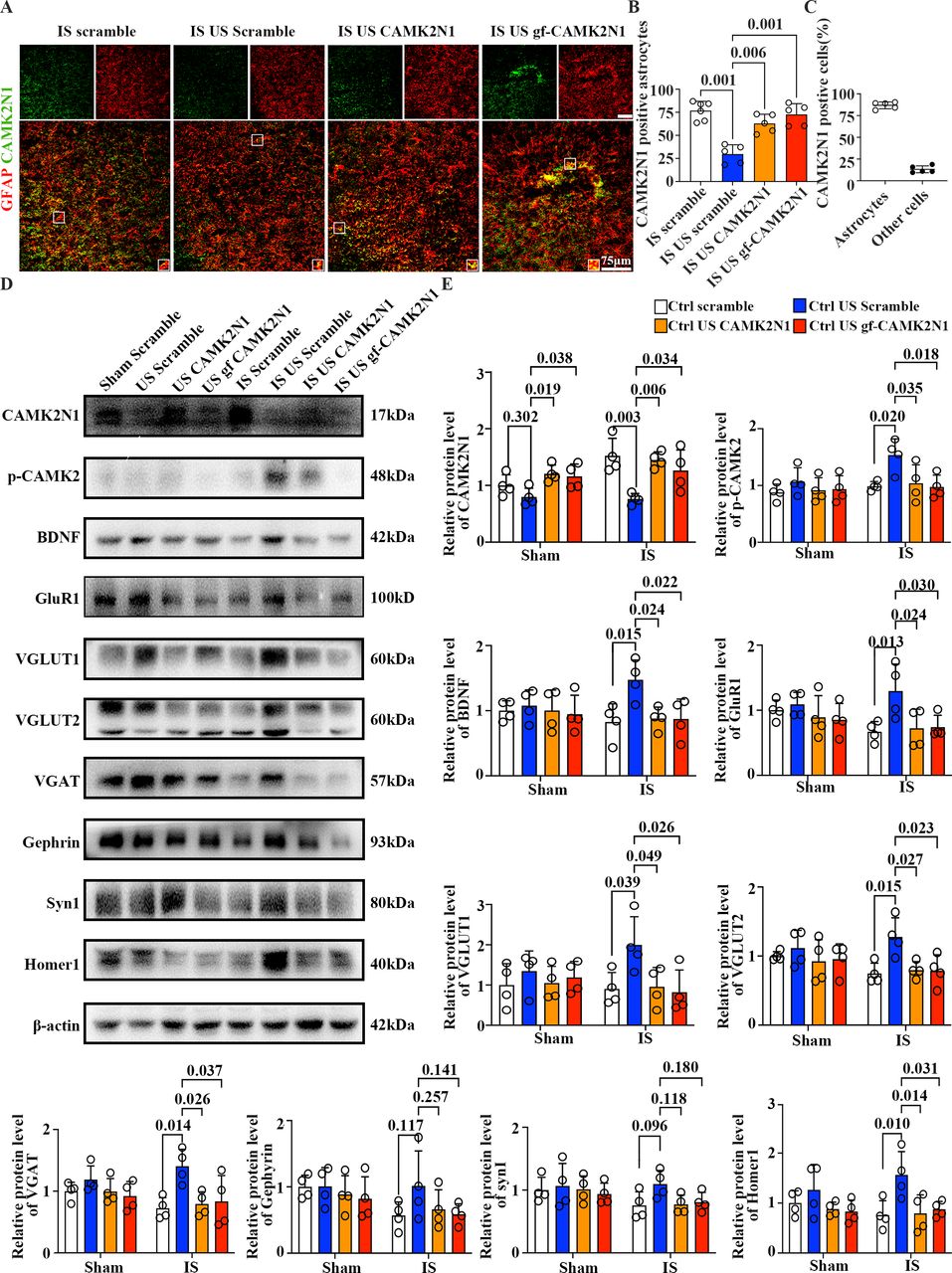
Astrocytic CAMK2N1 overexpression reversed the synapses increase after LIFUS in MCAO mice. (A) Representative images of CAMK2N1 (green) signals and GFAP+ astrocytes (red) in IS scramble, IS US scramble, IS US CAMK2N1, and IS US gf-CAMK2N1 mice. Scale bar = 75 µm. (B) Corresponding quantification of CAMK2N1 mRNA expression level in different groups and (C) percentile of astrocytes, (n= 3 mice/group). (D) Western blotting and (E) quantification of CAMK2N1, p-CAMK2, BDNF, GluR1, VGLUT1, VGLUT2, VGAT, Gephyrin, SynI, Homer1, respectively, from left to right and up to down, relative to β-actin and normalised to corresponding sham in ipsilateral hemisphere of mouse brain, (n=4 mice/group). Sham groups indicated sham scramble group, US group, US CAMK2N1 group and US gf-CAMK2N1 group. IS groups indicated IS scramble group, IS US scramble group, IS US sh (HMGB1) group and IS US gf-sh (HMGB1) group. Data are mean ± SD. IS, ischaemic stroke mice; IS US, ischaemic stroke mice treated with US; US, mice treated with ultrasound; LIFUS, low-intensity focused ultrasound stimulation.
- Figure 6
Astrocytic CAMK2N1 downregulated by LIFUS promoted electrical signals and increased dendritic spine density after LIFUS in MCAO mice. (A) Presentative Golgi staining images and quantitative analysis. Low magnification of Golgi staining images of neurons in the perifocal region of ipsilateral hemisphere. Scale bar=75 µm. (B) Representative images of dendritic spines and (C) a bar graph showed the number of total spines in the sham, US, US CAMK2N1, US gf-CAMK2N1, IS, IS US, IS US CAMK2N1, IS US gf-CAMK2N1 mice at 14 days after MCAO, (n=4 mice/group). (D) Average Ca2+ transients (detla F/F) and (E) variance of GCaMP6s signals were displayed at day 7 and day 13 after MCAO. (F) and (G) Heatmap displayed variance of the calcium activity of neurons during a 5-min records after ultrasound stimulation and CAMK2N1 overexpression respectively (n=3 mice/group). (H, I) Electromyography (EMG) records showed average EMG amplitude (detla A/A) and (J, K) variance heatmap during a 5-min records, (n=3 mice/group). (L) mNSS, (M) tail suspension, (N) grid walking (O) and rotarod test showed that neurobehavioural outcomes in different groups, (n=3 mice/group in the sham groups, n=12 mice/group in the IS groups). Ctrl (black line), Ctrl US (blue line), Ctrl US CAMK2N1 (yellow line), and Ctrl US gf-CAMK2N1 (red line) mice. Data are mean ± SD. IS, ischaemic stroke mice; IS US, ischaemic stroke mice treated with US; US, mice treated with ultrasound; LIFUS, low-intensity focused ultrasound stimulation.
- Figure 7
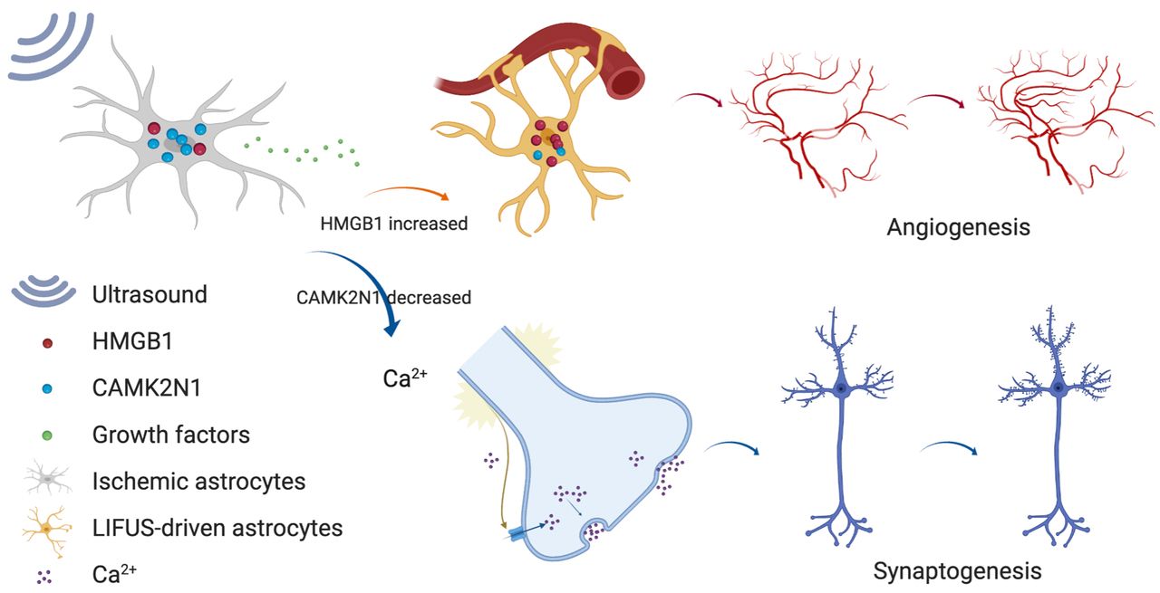
Low-intensity focused ultrasound stimulation promotes stroke recovery via astrocytic HMGB1 and CAMK2N1 in mice. LIFUS, low-intensity focused ultrasound stimulation.


Supplementary Materials
Supplementary data
Additional Files
Supplementary Data
This web only file has been produced by the BMJ Publishing Group from an electronic file supplied by the author(s) and has not been edited for content.


