
Main menu

  • Online first
    • Online first
  • Current issue
    • Current issue
  • Archive
    • Archive
  • Submit a paper
    • Online submission site
    • Instructions for authors
  • About the journal
    • About the journal
    • Editorial board
    • Instructions for authors
    • FAQs
    • Chinese Stroke Association
  • Help
    • Contact us
    • Feedback form
    • Reprints
    • Permissions
    • Advertising
  • BMJ Journals

User menu

  • Login

Search

  • Advanced search
  • BMJ Journals
  • Login
  • Facebook
  • Twitter
Stroke and Vascular Neurology

Advanced Search

  • Online first
    • Online first
  • Current issue
    • Current issue
  • Archive
    • Archive
  • Submit a paper
    • Online submission site
    • Instructions for authors
  • About the journal
    • About the journal
    • Editorial board
    • Instructions for authors
    • FAQs
    • Chinese Stroke Association
  • Help
    • Contact us
    • Feedback form
    • Reprints
    • Permissions
    • Advertising
Open Access

Altered excitability of motor neuron pathways after stroke: more than upper motor neuron impairments

Pingping Li, Chunyong Chen, Baozi Huang, Zimu Jiang, Jiating Wei, Jinsheng Zeng
DOI: 10.1136/svn-2022-001568 Published 20 December 2022
Pingping Li
1 Department of Neurology and Stroke Center, National Key Clinical Department and Key Discipline of Neurology, The First Affiliated Hospital, Sun Yat-sen University, Guangzhou, Guangdong, China
2 Department of Neurology, The First Affiliated Hospital of Guangxi Medical University, Nanning, Guangxi, China
  • Find this author on Google Scholar
  • Find this author on PubMed
  • Search for this author on this site
  • ORCID record for Pingping Li
Chunyong Chen
1 Department of Neurology and Stroke Center, National Key Clinical Department and Key Discipline of Neurology, The First Affiliated Hospital, Sun Yat-sen University, Guangzhou, Guangdong, China
2 Department of Neurology, The First Affiliated Hospital of Guangxi Medical University, Nanning, Guangxi, China
  • Find this author on Google Scholar
  • Find this author on PubMed
  • Search for this author on this site
  • ORCID record for Chunyong Chen
Baozi Huang
1 Department of Neurology and Stroke Center, National Key Clinical Department and Key Discipline of Neurology, The First Affiliated Hospital, Sun Yat-sen University, Guangzhou, Guangdong, China
2 Department of Neurology, The First Affiliated Hospital of Guangxi Medical University, Nanning, Guangxi, China
  • Find this author on Google Scholar
  • Find this author on PubMed
  • Search for this author on this site
Zimu Jiang
1 Department of Neurology and Stroke Center, National Key Clinical Department and Key Discipline of Neurology, The First Affiliated Hospital, Sun Yat-sen University, Guangzhou, Guangdong, China
  • Find this author on Google Scholar
  • Find this author on PubMed
  • Search for this author on this site
Jiating Wei
1 Department of Neurology and Stroke Center, National Key Clinical Department and Key Discipline of Neurology, The First Affiliated Hospital, Sun Yat-sen University, Guangzhou, Guangdong, China
  • Find this author on Google Scholar
  • Find this author on PubMed
  • Search for this author on this site
Jinsheng Zeng
1 Department of Neurology and Stroke Center, National Key Clinical Department and Key Discipline of Neurology, The First Affiliated Hospital, Sun Yat-sen University, Guangzhou, Guangdong, China
  • Find this author on Google Scholar
  • Find this author on PubMed
  • Search for this author on this site
  • ORCID record for Jinsheng Zeng
  • Article
  • Figures & Data
  • eLetters
  • Info & Metrics
  • PDF
Loading

Article Figures & Data

Figures

  • Supplementary Materials
  • Additional Files
  • Figure 1
    • Download figure
    • Open in new tab
    • Download powerpoint
    Figure 1

    Hill and Valley Task of cynomolgus monkeys.Note: A: Hill Task of right hand; B: Hill Task of left hand; C: Valley Task of right hand; D: Valley Task of left hand.

  • Figure 2
    • Download figure
    • Open in new tab
    • Download powerpoint
    Figure 2

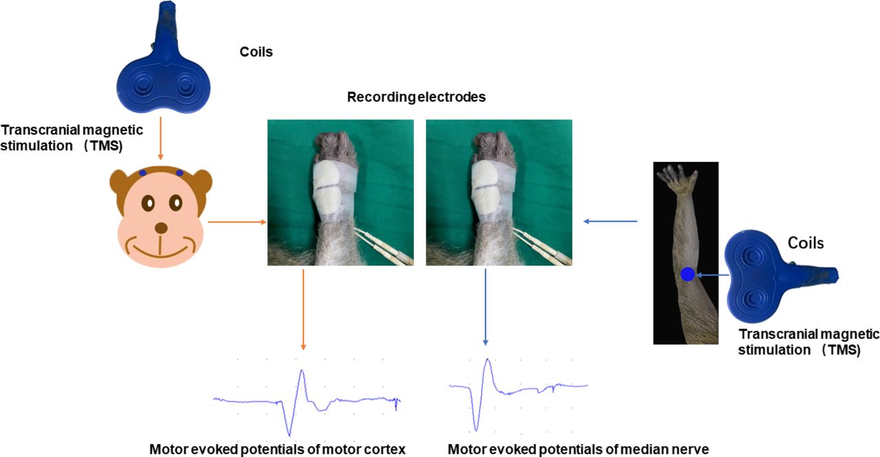
    Stimulation sites of magnetic stimulation and recording locations of motor evoked potentials.

  • Figure 3
    • Download figure
    • Open in new tab
    • Download powerpoint
    Figure 3

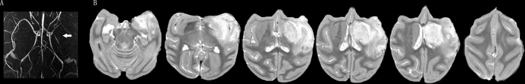
    MRA and T2-weighted MRI of cynomolgus monkeys 2 weeks after MCAO.Note: A: MRA indicated that the left middle cerebral artery was blocked (white arrow); B: high signal infarct lesion of left hemisphere was shown on the T2-weighted MRI.

  • Figure 4
    • Download figure
    • Open in new tab
    • Download powerpoint
    Figure 4

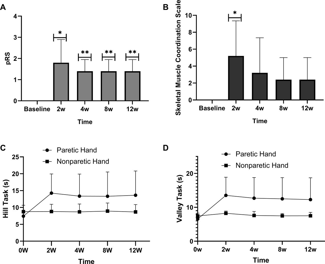
    pPS and motor function assessment.Note: A: pRS score; B: Skeletal muscle coordination scale score; C: Hill Task time; D: Valley Task time. * represents p value less than 0.05 compared with baseline; ** represents p value less than 0.001 compared with baseline. pRS, primate Rankin Scale.

  • Figure 5
    • Download figure
    • Open in new tab
    • Download powerpoint
    Figure 5

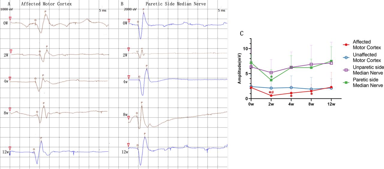
    MEP waveforms and amplitude dynamic evolution.Note: A+B: a single-onset MEP waveforms in a single monkey; A: MEP waveforms of the affected motor cortex; B: MEP waveforms of the paretic side median nerve. The red triangle indicated the stimulation start time. The length of each small square in A represented 5ms and the height represented 1000μV. The length of each small square in B represented 5ms and the height represented 2000μV. O indicated the MEP onset time and P represented the wave crest. Latency refers to the time from the onset of magnetic stimulation (red triangle) to the onset of MEP (O point), and amplitude refers to the longitudinal distance between the wave crest and the wave trough. C: Dynamic evolution of amplitude at different preoperative and postoperative time points.

  • Figure 6
    • Download figure
    • Open in new tab
    • Download powerpoint
    Figure 6

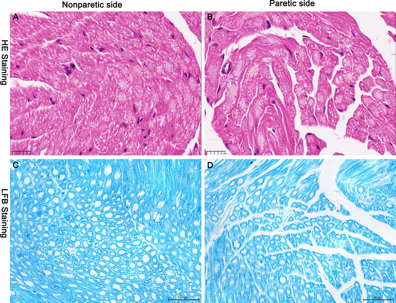
    HE staining and LFB staining of median nerve.Note: A+C: HE staining and LFB staining of nonparetic side median nerve with tight and regular arrangement of myelin sheaths; B+D: HE staining and LFB staining of paretic side median nerve with sparse and disorganized arrangement of myelin sheaths. LFB: LuxolFastBlue.

  • Figure 7
    • Download figure
    • Open in new tab
    • Download powerpoint
    Figure 7

    Immunofluorescence Double-labeling of Median Nerve.Note: In contrast to the nonparetic side median nerve, the MBP+ expression of the paretic side median nerve was less with disturbed arrangement of myelin structures.

  • Figure 8
    • Download figure
    • Open in new tab
    • Download powerpoint
    Figure 8

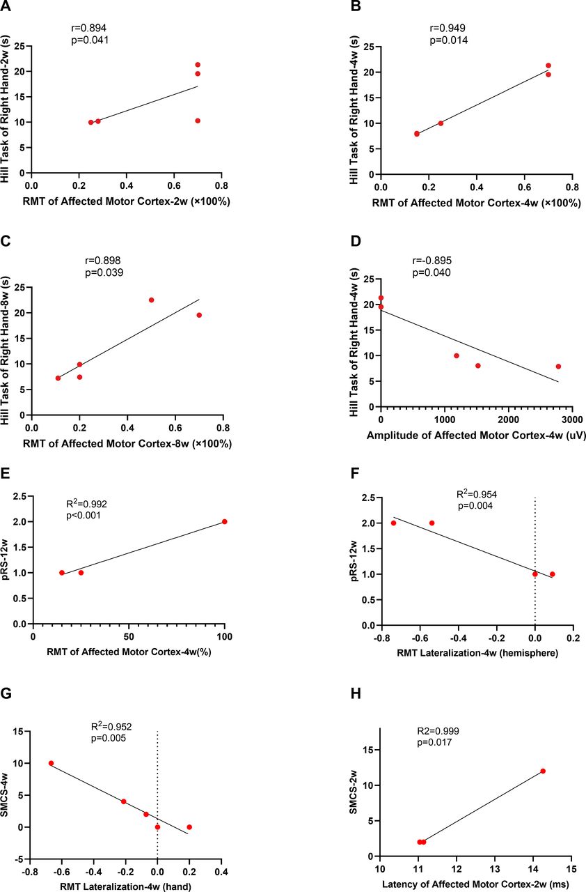
    Correlation analysis.Note: A: positive correlation between RMT of affected motor cortex and Hill Task of right hand at 2w; B: positive correlation between RMT of affected motor cortex and Hill Task of right hand at 4w; C: positive correlation between RMT of affected motor cortex and Hill Task of right hand at 8w; D: negative correlation between amplitude of affected motor cortex and Hill Task of right hand at 4w; E: automatic linear modeling of RMT of affected motor cortex (4w) and pRS (12w) with positive correlation; F: automatic linear modeling of RMT lateralization (4w) and pRS (12w) with negative correlation; G: automatic linear modelling of RMT lateralization (4w) and SMCS (4w) with negative correlation; H: automatic linear modeling of latency of affected motor cortex (2w) and SMCS (2w) with positive correlation. RMT, resting motor threshold. pRS, primate Rankin Scale. SMCS, Skeletal Muscle Coordination Scale.

Supplementary Materials

  • Figures
  • Additional Files
  • Supplementary data

    [svn-2022-001568supp001.pdf]

Additional Files

  • Figures
  • Supplementary Materials
  • Supplementary Data

    This web only file has been produced by the BMJ Publishing Group from an electronic file supplied by the author(s) and has not been edited for content.

    • Data supplement 1
PreviousNext
Back to top
Vol 7 Issue 6 Table of Contents
Stroke and Vascular Neurology: 7 (6)
  • Table of Contents
  • Table of Contents (PDF)
  • About the Cover
  • Index by author
  • Front Matter (PDF)
Email

Thank you for your interest in spreading the word on Stroke and Vascular Neurology.

NOTE: We only request your email address so that the person you are recommending the page to knows that you wanted them to see it, and that it is not junk mail. We do not capture any email address.

Enter multiple addresses on separate lines or separate them with commas.
Altered excitability of motor neuron pathways after stroke: more than upper motor neuron impairments
(Your Name) has sent you a message from Stroke and Vascular Neurology
(Your Name) thought you would like to see the Stroke and Vascular Neurology web site.
CAPTCHA
This question is for testing whether or not you are a human visitor and to prevent automated spam submissions.
Print
Alerts
Sign In to Email Alerts with your Email Address
Citation Tools
Altered excitability of motor neuron pathways after stroke: more than upper motor neuron impairments
Pingping Li, Chunyong Chen, Baozi Huang, Zimu Jiang, Jiating Wei, Jinsheng Zeng
Stroke and Vascular Neurology Dec 2022, 7 (6) 518-526; DOI: 10.1136/svn-2022-001568

Citation Manager Formats

  • BibTeX
  • Bookends
  • EasyBib
  • EndNote (tagged)
  • EndNote 8 (xml)
  • Medlars
  • Mendeley
  • Papers
  • RefWorks Tagged
  • Ref Manager
  • RIS
  • Zotero
Cite This
  • APA
  • Chicago
  • Endnote
  • MLA
Loading
Altered excitability of motor neuron pathways after stroke: more than upper motor neuron impairments
Pingping Li, Chunyong Chen, Baozi Huang, Zimu Jiang, Jiating Wei, Jinsheng Zeng
Stroke and Vascular Neurology Dec 2022, 7 (6) 518-526; DOI: 10.1136/svn-2022-001568
Download PDF

Share
Altered excitability of motor neuron pathways after stroke: more than upper motor neuron impairments
Pingping Li, Chunyong Chen, Baozi Huang, Zimu Jiang, Jiating Wei, Jinsheng Zeng
Stroke and Vascular Neurology Dec 2022, 7 (6) 518-526; DOI: 10.1136/svn-2022-001568
Reddit logo Twitter logo Facebook logo Mendeley logo
Respond to this article
  • Tweet Widget
  • Facebook Like
  • Google Plus One
  • Article
    • Abstract
    • Introduction
    • Materials and methods
    • Result
    • Discussion
    • Conclusions
    • Data availability statement
    • Ethics statements
    • Footnotes
    • References
  • Figures & Data
  • eLetters
  • Info & Metrics
  • PDF

Related Articles

Cited By...

More in this TOC Section

  • Stepwise improvement in intracerebral haematoma expansion prediction with advanced imaging: a comprehensive comparison of existing scores
  • Learning curve and embolisation strategy in single-stage surgery combined embolisation and microsurgery for brain arteriovenous malformations: results from a nationwide multicentre prospective registry study
  • Thrombus iodine-based perviousness is associated with recanalisation and functional outcomes in endovascular thrombectomy
Show more Original research

Similar Articles


CONTENT

  • Latest content
  • Current issue
  • Archive
  • eLetters
  • Sign up for email alerts
  • RSS

JOURNAL

  • About the journal
  • Editorial board
  • Recommend to librarian
  • Chinese Stroke Association

AUTHORS

  • Instructions for authors
  • Submit a paper
  • Track your article
  • Open Access at BMJ

HELP

  • Contact us
  • Reprints
  • Permissions
  • Advertising
  • Feedback form

© 2025 Chinese Stroke Association