

Article Figures & Data
Figures
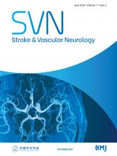
- Figure 1
Flow chart for patient inclusion.
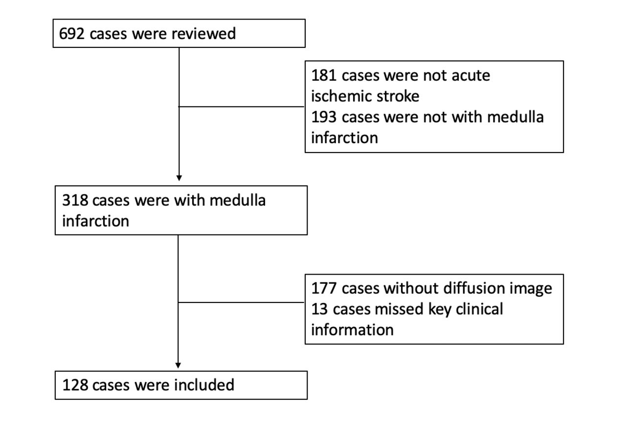
- Figure 2
Aetiologies of medullary infarction. Stroke aetiologies of medullary infarction (A); vertical and horizontal division of medulla (B); proportion of dissection in different lesion locations of medullary infarction vertically (C) and horizontally (D). AL, anterolateral; AM, anteromedial; C, caudal medulla; CE, cardiogenic embolism; L, lateral; LAA, large artery atherosclerosis; M, middle medulla; P, posterior; R, rostral medulla; SVO, small vessel occlusion.
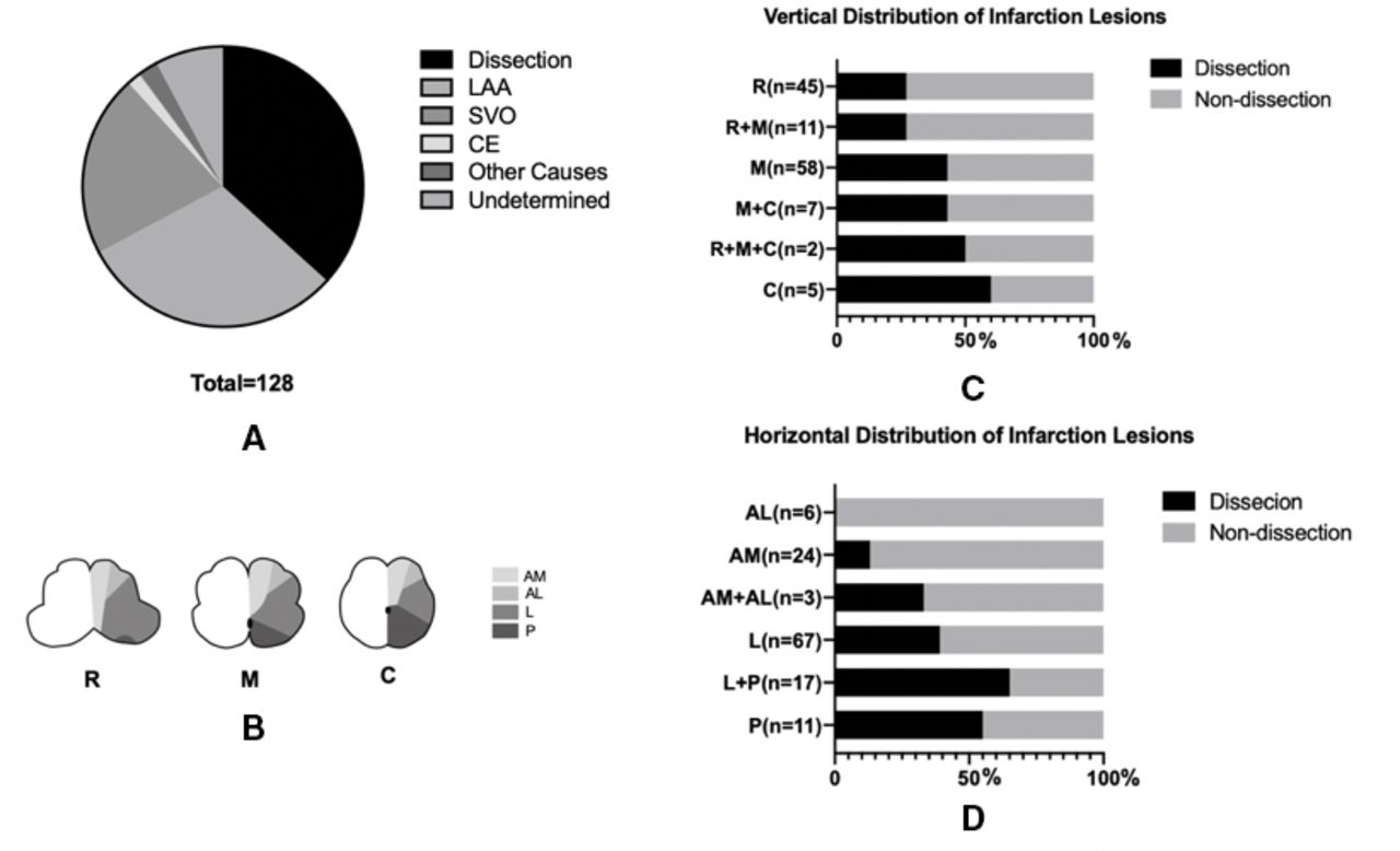
- Figure 3
Example of lesion topography of medullary infarction Medial MI (A) ; Lateral MI with ipsilateral hemiplegia (B); Avellis syndrome (C). CE, cardiogenic embolism; LAA, large artery atherosclerosis; MI, medullary infarction; SVO, small vessel occlusion.


Tables
- Table 1
Clinical features of MI caused by sVAD and non-sVAD
sVADN=47 Non-sVADN=81 P Sex, N (%) Female 10 (21.28) 12 (14.81) 0.350 Male 37 (78.72) 69 (85.19) Age, Med (IQR) 44 (34,53) 58 (52,66) 0.000 Onset, N (%) Sudden onset 34 (72.34) 73 (90.12) 0.009 Non-sudden onset 13 (27.66) 8 (9.87) Hypertension, N (%) 21 (44.68) 55 (67.90) 0.010 Coronary heart disease, N (%) 1 (2.13) 5 (6.17) 0.413 Diabetes, N (%) 9 (19.15) 37 (45.69) 0.003 Dyslipidaemia, N (%) 11 (23.40) 26 (32.10) 0.296 Atrial fibrillation, N (%) 1 (2.13) 2 (2.47) 1.000 Past stroke history, N (%) 1 (2.13) 10 (12.35) 0.054 Smoker, N (%) 20 (42.55) 27 (33.33) 0.297 Alcohol, N (%) 10 (21.28) 10 (12.35) 0.180 Family history of stroke, N (%) 1 (2.13) 5 (6.17) 0.157 Minor neck injury, N (%) 9 (19.15) 1 (1.23) 0.001 Headache, N (%) 22 (46.81) 6 (7.41) 0.000 Neck pain, N (%) 2 (4.26) 4 (4.94) 1.000 Side of infarction, N (%) Left 25 (53.19) 37 (45.68) 0.147 Right 22 (46.81) 38 (46.91) Bilateral 0 6 (7.41) Medial MI 3 (6.38) 30 (37.04) 0.000 Lateral MI 43 (91.48) 51 (62.96) 0.000 Both medial and lateral MI 1 (2.13) 0 0.367 Vertebral perforative infarction, N (%) 39 (82.98) 62 (76.54) 0.390 Multi-level medulla involvement, N (%) 8 (17.02) 19 (23.46) 0.390 Cerebellum involvement, N (%) 7 (14.89) 13 (16.05) 0.862 Involvement of other posterior vascular territories, N (%) 2 (4.26) 9 (11.11) 0.326 MI, medullary infarction; sVAD, spontaneous vertebral artery dissection.
- Table 2
Neurologic symptoms and signs of lateral MI
sVAD N=43 Non-sVAD N=51 P Ipsilateral facial paralysis, N (%) 4 (9.30) 11 (21.57) 0.157 Contralateral facial paralysis, N (%) 2 (4.65) 4 (7.84) 0.684 Bilateral facial paralysis, N (%) 0 1 (1.96) 1.000 Ipsilateral motor weakness, N (%) 8 (18.60) 11 (21.57) 0.721 Contralateral motor weakness, N (%) 2 (4.54) 5 (9.80) 0.448 Ipsilateral facial dysesthaesia, N (%) 23 (53.49) 19 (37.25) 0.115 Contralateral facial dysesthaesia, N (%) 5 (11.63) 4 (7.84) 0.727 Ipsilateral hemianesthaesia, N (%) 6 (13.95) 5 (9.80) 0.533 Contralateral hemianesthaesia, N (%) 30 (69.77) 24 (47.06) 0.027 Ataxia, N (%) 30 (69.77) 27 (52.94) 0.096 Dizziness, N (%) 27 (62.79) 29 (56.86) 0.560 Dysarthria, N (%) 16 (37.21) 20 (39.22) 0.842 Dysphagia, N (%) 27 (62.79) 27 (52.94) 0.336 Vertigo, N (%) 17 (39.53) 16 (31.37) 0.409 Nystagmus, N (%) 13 (30.23) 14 (27.45) 0.767 Nausea or vomiting, N (%) 27 (62.79) 24 (47.06) 0.127 MI, medullary infarction; sVAD, spontaneous vertebral artery dissection.
- Table 3
Vascular characteristics of sVAD in medial MI and lateral MI
Medial MI
N=3Lateral MI
N=43Both medial and lateral MI
N=1P Bilateral vertebral arteries dissection, N (%) 0 9 (20.93) 0 0.596 V4 segment involvement, N (%) 3 (100.00) 34 (79.07) 1 (100.00) 0.596 The site of artery dissection, N (%) 0.532 V1 0 2 (4.65) 0 V2 2 (66.67) 10 (23.26) 0 V3 1 (33.33) 15 (34.88) 0 V4 0 16 (37.21) 1 (100.00) Dissecting aneurysm, N (%) 0 2 (4.65) 0 0.907 Intimal flap, N (%) 0 2 (4.65) 0 0.907 Pearl-and-string sign, N (%) 1 (33.33) 6 (13.95) 0 0.604 Double-lumen, N (%) 1 (33.33) 4 (9.30) 0 0.401 Tapered steno-occlusion plus evidence of intramural haematoma, N (%) 1 (33.33) 29 (67.44) 1 (100.00) 0.372 MI, medullary infarction; sVAD, spontaneous vertebral artery dissection.
- Table 4
Independent factors associated with MI caused by sVAD
Factors Univariate analysis P Multivariable analysis P OR (95% CI) OR (95% CI) Age 0.907 (0.873 to 0.943) 0.000 0.935 (0.892 to 0.981) 0.006 Non-sudden onset 3.489 (1.322 to 9.206) 0.012 3.507 (1.060 to 11.599) 0.040 Hypertension 0.382 (0.182 to 0.801) 0.011 1.085 (0.370 to 3.179) 0.882 Diabetes 0.282 (0.121 to 0.658) 0.003 0.559 (0.187 to 1.669) 0.297 Minor neck injury 18.947 (2.316 to 155.00) 0.006 2.653 (0.264 to 26.676) 0.408 Headache 11.000 (4.007 to 30.196) 0.000 5.426 (1.673 to 17.599) 0.005 Lateral MI 6.665 (2.178 to 20.391) 0.001 2.477 (0.674 to 9.108) 0.172 MI, medullary infarction; sVAD, spontaneous vertebral artery dissection.


