

Article Figures & Data
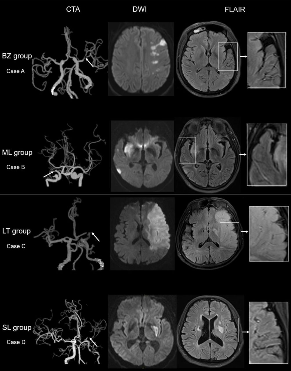
- Figure 1
Illustrative cases according to the DWI lesion patterns. Case A (BZ group) shows severe stenosis of left MCA on CTA, a border-zone infarct lesion on DWI and with FVH positive in FLAIR; FVHs located beyond the lesions. Case B (ML group) shows occlusion of the right MCA, two lesions in different artery territories on DWI and with FVH positive on FLAIR; FVHs located beyond the lesions. Case C (LT group) shows occlusion of the left MCA on CTA, a large territory infarct lesion on DWI and with FVH positive on FLAIR; FVHs located within or very closed to the lesions. Case D (SL group) shows near occlusion of the left MCA on CTA, a lesion in the basal ganglia on DWI and with FVH positive on FLAIR; FVHs located beyond the lesions. BZ group, border-zone infarct group; CTA, CT angiography; DWI, diffusion-weighted imaging; FLAIR, fluid-attenuated inversion recovery; FVH, FLAIR vascular hyperintensity; LT group, large territory infarct group; MCA, middle cerebral artery; ML group, multiple lesion infarcts group; SL group, single cortical or subcortical lesion group.
- Figure 2
The flow chart of patients included. BZ group, border-zone infarct group; DWI, diffusion-weighted imaging; LT group, large territory infarct group; MCA, middle cerebral artery; ML group, multiple lesion infarcts group; r-tPA, recombinant tissue plasminogen activator; SL group, single cortical or subcortical lesion group.


- Table 1
Clinical and demographic characteristics of FVH (+) and FVH (−) patients in all patients and four subgroups
All patients BZ group ML group LT group SL group FVH− FVH+ P value FVH− FVH+ P value FVH− FVH+ P value FVH− FVH+ P value FVH− FVH+ P value N=103 (%) N=100 (%) N=43 (%) N=29 (%) N=25 (%) N=39 (%) N=11 (%) N=26 (%) N=24 (%) N=6 (%) Sex (men) 82 (79.6) 87 (87.0) 0.159 35 (81.4) 25 (86.2) 0.830 21 (84.0) 33 (84.6) 0.999 9 (81.8) 23 (88.5) 0.989 17 (70.8) 6 (100) 0.331 Age 63.2±10.1 63.4±10.4 0.870 64.7±9.4 63.6±10.1 0.634 62.7±9.5 63.3±10.2 0.826 66.6±12.7 62.9±11.6 0.395 59.3±10.1 65.7±9.8 0.174 Hypertension 81 (78.6) 72 (72) 0.272 34 (79.1) 19 (65.5) 0.201 20 (80.0) 28 (71.8) 0.460 7 (63.6) 21 (80.8) 0.490 20 (83.3) 4 (66.7) 0.732 Diabetes mellitus 34 (33.0) 36 (36.0) 0.654 17 (39.5) 12 (41.4) 0.876 6 (24.0) 11 (28.2) 0.710 2 (18.2) 10 (38.5) 0.412 9 (37.5) 3 (50.0) 0.926 Coronary heart disease 14 (13.6) 19 (19.0) 0.296 6 (14.0) 6 (20.7) 0.667 2 (8.0) 7 (17.9) 0.454 3 (27.3) 6 (23.1) 0.999 3 (12.5) 0 (0) 0.999 Hyperlipidaemia 55 (53.4) 42 (42.0) 0.104 25 (58.1) 15 (51.7) 0.591 14 (56.0) 17 (43.6) 0.332 4 (36.4) 9 (34.6) 0.999 12 (50.0) 1 (16.7) 0.311 Previous stroke 29 (28.2) 25 (25.0) 0.611 11 (25.6) 8 (27.6) 0.850 10 (40.0) 7 (17.9) 0.051 3 (27.3) 5 (19.2) 0.915 5 (20.8) 5 (83.3) 0.015* Smoking 58 (56.3) 62 (62.0) 0.410 24 (55.8) 17 (58.6) 0.814 15 (60.0) 23 (59.0) 0.935 8 (72.7) 19 (73.1) 0.999 11 (45.8) 3 (50.0) 0.999 Drinking 57 (55.3) 54 (54.0) 0.848 24 (55.8) 15 (51.7) 0.733 13 (52.0) 25 (64.1) 0.336 7 (63.6) 13 (50.0) 0.447 13 (54.2) 1 (16.7) 0.234 Lacunar infarct 56 (54.4) 62 (62.0) 0.271 23 (53.5) 18 (62.1) 0.471 16 (64.0) 26 (66.7) 0.827 4 (36.4) 15 (57.7) 0.235 13 (54.2) 3 (50.0) 0.999 Occlusion 11 (10.7) 58 (58.0) <0.001* 2 (4.7) 13 (44.8) <0.001* 5 (20.0) 24 (61.5) 0.001* 3 (27.3) 17 (65.4) 0.033* 1 (4.2) 4 (66.7) 0.002* WMH (subcortical) 0 (0–1) 1 (0–1) 0.366 1 (0–1) 0 (0–1) 0.643 0 (0–1) 1 (0–2) 0.265 0 (0–1) 0 (0–1) 0.750 0 (0–1) 1 (0–2.25) 0.209 WMH (periventricular) 1 (0–2) 1 (0–2) 0.992 1 (1–2) 1 (0–1.5) 0.471 1 (0.5–1.5) 1 (1–3) 0.295 1 (0–2) 1 (0–1) 0.559 1 (0.25–2) 1 (1–1.25) 0.732 NIHSS-ad 5 (3–9) 7.5 (4–12) 0.002* 5 (3–9) 6 (4–10.5) 0.149 5 (4–9.5) 5 (4–11) 0.830 8.7±6.0 11.1±5.1 0.224 5.0±2.4 7.2±2.3 0.064 mRS ≥2 36 (34.3) 69 (65.7) <0.001* 10 (23.3) 21 (72.4) <0.001* 8 (32.0) 24 (61.5) 0.021* 6 (54.5) 22 (84.6) 0.126 12 (50.0) 1 (16.7) 0.311 mRS ≥3 18 (17.5) 47 (47.0) <0.001* 3 (7.0) 14 (48.3) <0.001* 4 (16.0) 15 (38.5) 0.055 4 (36.4) 18 (69.2) 0.135 7 (29.2) 0 (0) 0.331 mRS at discharge 1 (1–2) 2 (1–4) <0.001* 1 (0–1) 2 (1–4) <0.001* 1 (1–2) 2 (1–4) 0.011* 2 (1–4) 4 (2–4) 0.088 1.5 (1–3) 1 (1–1.25) 0.210 Hospitalisation (days) 7 (6–9) 8 (7–10) 0.009* 7 (6–8) 8 (7–10) 0.014* 7 (6–8) 9 (7–10) 0.007* 10(9-11) 9 (6.75–11.25) 0.277 8 (6.25–10) 7.5 (6.5–8.5) 0.529 r-tPA to MRI time (days) 2 (1–3) 2 (1–2) 0.624 2 (1–3) 2 (1–3) 0.915 2 (1–3) 1 (1–3) 0.057 2 (1–4) 2 (1–2) 0.180 1 (1–2) 1.5 (1–2.25) 0.668 Data are presented as median (IQR), mean±SD or n (%).
*Significantly different.
BZ group, border-zone infarct group; FVH, fluid-attenuated inversion recovery vascular hyperintensity; LT group, large territory infarct group; ML group, multiple lesion infarcts group; mRS, modified Rankin Scale; NIHSS-ad, National Institute of Healthy Stroke Scale at admission; r-tPA, recombinant tissue plasminogen activator; SL group, single cortical or subcortical lesion group; WMH, white matter hyperintensity.
- Table 2
Results of multiple logistic regression analysis for unfavourable (mRS ≥2) and poor (mRS ≥3) outcome in all patients and four subgroups
All patients BZ group ML group LT group SL group OR (95% CI) P value OR (95% CI) P value OR (95% CI) P value OR (95% CI) P value OR (95% CI) P value Model 1, mRS ≥2 FVH (+) 3.02 (1.49 to 6.13) 0.002 4.22 (1.25 to 14.25) 0.021 5.44 (1.41 to 20.92) 0.014 1.95 (0.24 to 15.90) 0.531 0.24 (0.01 to 7.36) 0.410 Male 2.00 (0.86 to 4.66) 0.110 0.20 (0.02 to 2.12) 0.180 0.91 (0.17 to 4.99) 0.911 8.74 (0.31 to 246.91) 0.203 3.55 (0.49 to 25.78) 0.211 Age 1.00 (0.97 to 1.03) 0.870 1.01 (0.96 to 1.08) 0.654 1.02 (0.96 to 1.09) 0.448 0.97 (0.89 to 1.06) 0.499 1.01 (0.93 to 1.11) 0.761 Occlusion 1.67 (0.77 to 3.59) 0.192 1.14 (0.97 to 1.33) 0.103 0.58 (0.15 to 2.18) 0.420 20.51 (1.69 to 248.68) 0.018 1.08 (0.03 to 33.89) 0.965 NIHSS-ad 1.18 (1.09 to 1.28) <0.001 14.19 (1.40 to 143.85) 0.025 1.24 (1.06 to 1.46) 0.007 1.24 (0.96 to 1.60) 0.093 1.03 (0.72 to 1.46) 0.882 Model 2, mRS ≥3 FVH (+) 2.25 (1.01 to 4.97) 0.046 5.52 (0.98 to 31.07) 0.053 4.09 (1.04 to 16.16) 0.045 1.53 (0.16 to 14.40) 0.709 NA NA Male 1.40 (0.54 to 3.62) 0.488 0.30 (0.02 to 3.94) 0.363 0.56 (0.09 to 3.58) 0.537 8.48 (0.33 to 217.26) 0.197 0.70 (0.08 to 6.26) 0.754 Age 1.00 (0.97 to 1.03) 0.937 1.05 (0.96 to 1.14) 0.338 1.03 (0.97 to 1.09) 0.318 0.88 (0.77 to 1.00) 0.051 1.04 (0.93 to 1.16) 0.511 Occlusion 2.90 (1.32 to 6.37) 0.008 32.43 (3.21 to 327.27) 0.003 2.10 (0.57 to 7.82) 0.268 5.39 (0.54 to 54.04) 0.152 0.01 (0.01 to 999.99) 0.999 NIHSS-ad 1.21 (1.11 to 1.31) <0.001 1.34 (1.07 to 1.68) 0.010 1.14 (1.02 to 1.27) 0.019 1.69 (1.18 to 2.44) 0.004 1.30 (0.83 to 2.02) 0.253 Model 1 used mRS ≥2 as dependent variable; model 2 used mRS ≥3 as dependent variable.
The bolded entries mean significance.
BZ group, border-zone infarct group; FVH, fluid-attenuated inversion recovery vascular hyperintensity; LT group, large territory infarct group; ML group, multiple lesion infarcts group; mRS, modified Rankin Scale; NA, not available; NIHSS-ad, National Institute of Healthy Stroke Scale at admission; SL group, single cortical or subcortical lesion group.
- Table 3
The distribution of FVHs in four subgroups
FVH within DWI lesion FVH beyond DWI lesion FVH (both within and beyond) Total patients, n=100, (%) 14 (14.0) 73 (73.0) 13 (13.0) BZ group, n=29, (%) 1 (3.4) 26 (89.7) 2 (6.9) ML group, n=39, (%) 0 (0) 38 (97.4) 1 (2.6) LT group, n=26, (%) 13 (50.0) 3 (11.5) 10 (38.5) SL group, n=6, (%) 0 (0) 6 (100) 0 (0) BZ group, border-zone infarct group; DWI, diffusion-weighted imaging; FVH, fluid-attenuated inversion recovery vascular hyperintensity; LT group, large territory infarct group; ML group, multiple lesion infarcts group; SL group, single cortical or subcortical lesion group.
Supplementary Materials
Supplementary data
Additional Files
Supplementary Data
This web only file has been produced by the BMJ Publishing Group from an electronic file supplied by the author(s) and has not been edited for content.


