

Abstract
Background Given that associations of Life’s Essential 8 (LE8) and cerebral small vessel disease (CSVD) or its imaging markers were unclear, we examined relationship between them.
Methods The cross-sectional study included community residents from the PolyvasculaR Evaluation for Cognitive Impairment and vaScular Events study. We calculated the total LE8 score, medical LE8 score and behavioural score, and categorised them into low (<60), moderate (60–79) or high (≥80) group. MRI markers included lacunes, white matter hyperintensities (WMH), enlarged perivascular spaces in basal ganglia (BG-EPVS) and cerebral microbleeds (CMB). In respect of, total CSVD score (0–4 points), WMH, lacunes or CMB were categorised as two grades, and BG-EPVS (N>10) was allocated one point. Based on modified total CSVD score (0–6 points), WMH or CMB was modified to three grades, and BG-EPVS (N>20) was allocated one point.
Results Among 3061 participants in this study, 1424 (46.5%) were male. Higher LE8 score was associated with lower total CSVD score (moderate vs low: cOR 0.78, 95% CI 0.63 to 0.96; high vs low: cOR 0.44, 95% CI 0.33 to 0.59), and the medical score was inversely related to the total CSVD score. Furthermore, the medical score was inversely related to odds of WMH (p<0.05), modified WMH (p<0.05), lacunes (p<0.05) or BG-EPVS (p<0.05), and the behavioural score were inversely related to the odds of lacunes and BG-EPVS.
Conclusions Higher LE8 score which indicates better cardiovascular status was associated with lower burden of CSVD and its MRI markers. Longitudinal studies are needed to examine the causality.
WHAT IS ALREADY KNOWN ON THIS TOPIC
Some individual lifestyle factors were associated with cerebral small vessel disease (CSVD) or its MRI markers, but related results were not consistent. The score of cardiovascular health has been put forward as a tool for promoting brain health.
WHAT THIS STUDY ADDS
This study explored whether Life’s Essential 8 (LE8) score, which is a novel score to evaluate cardiovascular health, was associated with CSVD and is MRI markers.
HOW THIS STUDY MIGHT AFFECT RESEARCH, PRACTICE OR POLICY
Findings of this study provided clues for future studies to identify the causal relationship between LE8 and CSVD and provide people with an index to evaluate the status of cerebral small vascular health.
Introduction
Cerebral small vessel disease (CSVD) encompasses a group of pathological processes that affect small penetrating vessels (arterioles, capillaries and venules) of the brain.1 It is a serious threat to public health and poses substantial burden on patients, their proxies, societies and healthcare systems.2 Previous studies suggested some individual lifestyle risk factors were associated with CSVD, but related results were not consistent.3 4 The mechanisms associating these risk factors and CSVD pathology are still not fully understood.
In 2022, the American Heart Association proposed an overall score—Life’s Essential 8 (LE8)—which is an enhanced approach to assessing cardiovascular health and updated from Life’s Simple 7 (LS7). LE8, which assessed cardiovascular health status, was an effective means to monitor public and individual health and a strong indicator of the extraordinary potential of primordial prevention strategies to improve and extend countless lives.5 The score of cardiovascular health has been put forward as a tool for promoting brain health.6 Previous studies examined the associations between LS7 and stroke or dementia which is closely related to CSVD,7 8 whereas few studies investigated the associations between these cardiovascular health scores and CSVD, especially for the novel LE8 score. The association between LE8 and CSVD or its MRI markers was unclear.
In this study, we investigated associations of LE8 with presence of CSVD, total CSVD burden and imaging features in community-dwelling adults based on the PolyvasculaR Evaluation for Cognitive Impairment and vaScular Events (PRECISE) study.
Method
Study design
This study was based on the PRECISE study, a population-based prospective cohort study.9 Briefly, 3067 older adults (50–75 years) were selected from 6 villages and 4 communities via cluster sampling between 2017 and 2019. Participants were excluded if they met any of the following criteria: with contraindications to MRI or CT angiography, life expectancy ≤4 years due to advanced cancers and other diseases or mental diseases.9 Our cross-sectional study was conducted among participants with complete baseline information of LE8, CSVD and MRI markers in the PRECISE study. This study was reported following the ‘Strengthening the Reporting of Observational Studies in Epidemiology’ guidelines.10
Data collection
In baseline, trained researchers collected information on demographics, medical history, routine physical examination, concomitant medication and lifestyle factors according to a case report form. Body mass index (BMI) was equal to the weight in kilograms divided by height in metres squared. The blood pressure was measured three times and recorded as the average of the second and third measurements. In addition, we collected fasting blood samples for participants and conducted a blood routine examination.
Life’s Essential 8
The components of LE8 included four medical metrics (BMI, blood lipids, blood glucose and blood pressure) and four behavioural metrics (diet, physical activity, nicotine exposure and sleep). Each metric was evaluated from 0 to 100 points, which meant from lower to higher health conditions of lifestyle. The LE8 score, medical score or behavioural score was equal to the unweighted average of all components which it includes, and the higher points indicated the healthier lifestyle.5 Furthermore, the LE8 score, medical score or behavioural score was categorised as low (0–60), moderate (60–80) or high (80–100) group. The details of scoring are presented in online supplemental table S1.
Supplementary data
Outcomes measurements
MRIs were acquired through a 3.0T MRI scanner (Ingenia 3.0T, Philips, Best, The Netherlands) by trained researchers in accordance with a standardised protocol. The MRI sequences included three-dimensional T1-weighted magnetisation-prepared rapid-acquisition gradient-echo, axial T2-weighted, fluid-attenuated inversion recovery and axial susceptibility-weighted imaging in online supplemental table S2. The researchers collected the imaging data in digital imaging and communications in medicine format on discs and sent it to the imaging research centre at Beijing Tiantan Hospital for analysis.
We defined MRI markers of CSVD according to the Standards for Reporting Vascular Changes on Neuroimaging criteria.11 White matter hyperintensities (WMH) are areas of signal abnormality in the brain white matter that show up increased brightness on T2-weighted images.12 The periventricular WMH (PV-WMH) and deep-WMH were evaluated by the Fazekas rating scale.12 Lacunes are rounded or ovoid, subcortical, fluid-filled cavities (3–15 mm in diameter) whose signals are similar to CSF.11 Cerebral microbleeds (CMBs) are small areas (2–10 mm in diameter) of the signal void with associated blooming which are seen on T2-weighted MRI.11 Enlarged perivascular spaces (EPVS) are fluid-filled spaces that follow the typical course of a vessel as it goes through grey or white matter.11 EPVS in the basal ganglia (BG-EPVS) was graded with the semiquantitative rating scale designed by the Edinburg group.13 The MRI markers were evaluated by four skilled raters (M Zhou, YC, J Pi, M Zhao) with one rater evaluating two markers. They were blinded to participants’ information. An additional senior neurologist (YY) who was unaware of initial results made the ultimate assessment of images with inconsistent results. Kappa coefficients between raters were as follows: 0.82 (Fazekas scale of WMH), 0.80 (the presence of lacune), 0.90 (the severity of EPVS) and 0.80 (the presence of CMB).
Total CSVD score14 and modified total CSVD score15 were used to assess CSVD and MRI markers, which include WMH, lacunes, CMBs and BG-EPVS. The total CSVD score designed by Wardlaw’s group was 0–4 points, and a higher score indicates a higher burden of CSVD. The details of it are as follows14: one point was awarded to the present of lacunes or CMBs; one point was awarded to moderate to severe (>10) BG-EPVS, one point was awarded to either confluent deep WMH (Fazekas scale 2 or 3) or irregular PV-WMH extending into the deep white matter (Fazekas score 3). The presence of CSVD (Wardlaw) was defined as total CSVD score ≥1. The modified CSVD score developed by Rothwell’s group was 0–6 points, and a higher score means a higher burden of CSVD. The details of the modified total CSVD score developed by Rothwell’s group was 0–6 points, and a higher score means a higher burden of CSVD. The details of it are as follows15: one point was allocated to the moderate degree of WMH (combined periventricular and subcortical WMH score of 3–4), presence of lacunes, 1–4 CMBs or frequent to severe (N>20) BG-EPVS; two points were allocated to the severe WMH (combined periventricular and subcortical WMH score of 5–6) or ≥5 microbleeds. The presence of CSVD (Rothwell) was defined as a modified total CSVD score≥1.
Statistical methods
For descriptive analysis, continuous variables are presented as mean with SD or median with IQR, while categorical variables are presented as frequencies with percentages. Comparisons of baseline characteristics between two groups were performed with the χ2 test, Fisher’s exact test, Student’s t-test or Mann-Whitney U test. We examined the associations of LE8 and its subscales with the presence of CSVD by using the binary logistic regression models and calculated the ORs and 95% CIs. Furthermore, we examined the associations of LE8 and its subscales with WMH, lacunes, CMBs, BG-EPVS and modified BG-EPVS through the same models. For the associations of LE8 and its subscale with total CSVD score, modified total CSVD score, modified WMH and modified CMBs, the ordinal logistic regression models were performed and the common ORs (cOR) with 95% CIs were calculated. Two models were conducted for the presence of CSVD, total CSVD score and MRI markers. In model 1, we performed a univariable analysis. In model 2, we performed a multivariable analysis with adjustments for the covariates including age, sex, estimated glomerular filtration rate and history of antiplatelet and anticoagulants drugs. All analyses were performed by using SAS software V.9.4 (SAS Institute). A p<0.05 was considered statistically significant.
Result
Among the 3067 participants, data on all LE8 metrics and MRIs of CSVD were available for 3061 participants. The mean age of 3061 participants was 61.2 (SD 6.7 years), and 1424 (46.5%) were male. In addition, 934 (30.5%) individuals presented with CSVD (Wardlaw), while 41.5% with CSVD (Rothwell). As presented in table 1, the participants with CSVD were older, more likely to be male and had a higher prevalence of hypertension and diabetes than those without CSVD.
Baseline characteristics for presence or absence of CSVD
The proportions of presence of CSVD by categories of LE8 are shown in table 2. The presence of CSVD (Wardlaw) in low, moderate and high LE8 score group were 37.7%, 32.2% and 18.6%, while the presence of CSVD (Rothwell) were 48.4%, 42.9% and 31.0%, respectively. After adjustment for covariates, participants in the high LE8 score group had a lower odd of presence of CSVD (Wardlaw) (OR 0.50, 95% CI 0.37 to 0.67) or CSVD (Rothwell) (OR 0.59, 95% CI 0.45 to 0.78). Focusing on the subscales, higher medical score was associated with lower odds of presence of CSVD (Wardlaw, OR 0.53, 95% CI 0.42 to 0.66; Rothwell, OR 0.63, 95% CI 0.51 to 0.78). However, there was no significant difference in the presence of CSVD between participants with different behavioural subscale score.
ORs for presence or absence of CSVD according to the LE8 score and its subscales
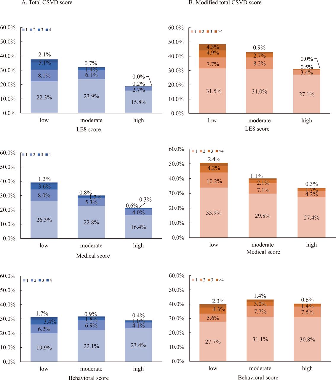
The distribution of total CSVD score and modified total CSVD score in different LE8 and its subscales categories are shown in figure 1. After adjusting for potential covariates, for the LE8 score, both moderate (cOR 0.78, 95% CI 0.63 to 0.96) and high (cOR 0.44, 95% CI 0.33 to 0.59) score categories were associated with lower odds of total CSVD score (figure 2). The associations between the LE8 score and modified total CSVD score were also significant. In further analyses of medical and behavioural subscales, the medical score was inversely associated with the total CSVD score or modified total CSVD score, while the behavioural subscale was not (figure 2).

Proportions of total CSVD score according to LE8 score and its subscales. LE8 score, medical score or behavioural score was categorised as low (0–60), moderate (60–80) or high (80–100) group. CSVD, cerebral small vessel disease; LE8, Life’s Essential 8.

Common ORs for total CSVD score according to LE8 score and its subscales. Common ORs for total CSVD score according to LE8 score categories adjusted for covariates in model 2. LE8 score, medical score or behavioural score was categorised as low (0–60), moderate (60–80) or high (80–100) group. CSVD, cerebral small vessel disease; LE8, Life’s Essential 8.
The associations between LE8 or its subscales and MRI markers are presented in table 3. LE8 and medical scores were inversely related to WMH and lacunes based on two criteria. The high behavioural score category was associated with lower odds of lacunes (OR 0.47, 95% CI 0.29 to 0.77). In addition, the high LE8 score category was associated with lower odds of CMB (OR 0.62, 95% CI 0.40 to 0.97) or modified CMB (cOR 0.61, 95% CI 0.39 to 0.95). Furthermore, LE8 score and its subscales scores were inversely associated with BG-EPVS.
OR for MRI markers based on the LE8 score and its subscales
Discussion
In the community-based study, our results indicated that higher LE8 and medical scores were related to lower odds of the presence of CSVD, total CSVD score and modified total CSVD score. Furthermore, among the MRI markers, inverse relationships were found between LE8 score and odds of each marker except for modified BG-EPVS. Inverse relationships were found between medical score and odds of WMH, modified WMH, lacunes, or BG-EPVS and between behavioural score and odds of lacunes or BG-EPVS.
To our knowledge, most studies reported that LS7, vascular risk factors or behavioural risk factors were associated with stroke or CSVD.3 16 17 Previous studies have suggested that the LS7 score was inversely associated with risks of stroke.18–20 A study based on community population has reported that LS7 was inversely associated with the risk of CSVD.17 These findings were consistent with our results on associations of LE8 and medical scores with presence of CSVD and total CSVD score. Compared with LS7, the LE8 was updated in scoring for all metrics and in assessing diet, smoking and blood glucose. In addition, researchers added a new metric—sleep—into LE8.5 A review focusing on Alzheimer’s disease and the blood–brain barrier suggested that sleep plays a crucial role in resistance to CSVD.21 In parallel, previous studies reported the relationships between some individual risk factors and the risk of MRI markers. Hypertension, hyperlipidaemia, diabetes or BMI was associated with the risk of WMH, while hypertension or diabetes was associated with the risk of lacunes.4 22–24 In addition, hypertension was related to CMB.25 This evidence supported our findings that the medical score was inversely related to the risk of MRI markers. Moreover, several studies have examined the association between some lifestyles and MRI markers which suggested that the amount or type of activity and dietary patterns were associated with WMH or lacunes, between the type of activity and WMH.26–28 In other studies, smoking and sleep were associated with the risk of WMH, CMB or EPVS.25 29 30 These findings provided a basis for the result that behavioural score was associated with the risk of lacunes or BG-EPVS. Overall, as the method of reducing the risk of CSVD was controversial and the importance of risk factors clustering was recognised,3 31 our observational study assessed overall lifestyle risk of CSVD using the comprehensive score—LE8 and provided an index which may help to evaluate the state of cerebral small vascular health. Future studies should be considered to examine whether improving the modifiable risk factors can reduce the risk of CSVD.
The underlying mechanisms for the association between LE8 and CSVD are not identified. There are several hypothetical mechanisms. Previous studies shown that hypertension may be indirectly related to WMH progression via arterial compliance,32 and the underlying pathophysiological process between hypertension and CMB may be a small vessel arteriopathy with changes of lipohyalinosis.25 In parallel, hyperlipidaemia leads to microvascular haemodynamic regulation disorder, which increases viscosity and resistance of blood flow, for the progression of WMH.4 Meanwhile, visceral obesity contributed to deep WMH through increases in proinflammatory cytokines.24 In addition, the potential explanation between a dietary pattern and MRI markers may be related to improved endothelial function, adiposity and lower levels of inflammatory markers.28 33 Furthermore, poor sleep efficiency was independently associated with BG-EPVS according to altering waste clearance mechanisms,30 and sleep may be associated with MRI markers according to inflammation.34
This study has several limitations. First, our study was a cross-sectional observational study, and the causal relationship between LE8 and CSVD cannot be established. As the PRECISE study is currently ongoing, we may investigate the causal relationship with longitudinal follow-up data. Second, the assessment of diet was according to the Mediterranean Eating Pattern for Americans (MEPA). In our study, we collected the information of green leafy vegetables, fruit, meat, fish, poultry and alcohol, other data of the MEPA was missing, thus we may underestimate the diet score of participants. Third, most of the participants in the PRECISE study were from rural areas. They work in hard physical work, so their level of physical activity may be higher than people in urban areas. Future studies with the population in urban or larger sample sizes are needed to validate our findings.
Conclusion
In conclusion, the higher LE8 and its subscale scores, indicating healthier lifestyle, were associated with lower odds of CSVD. The medical score was inversely associated with the odds of WMH, modified WMH, lacunes or BG-EPVS. Meanwhile, the higher behavioural score was associated with lower odds of lacunes or BG-EPVS. Prospective studies are needed to validate these results and to examine the relationship between LE8 and the progress of CSVD.
Data availability statement
Data are available on reasonable request. The data are available to researchers on reasonable request from the corresponding author.
Ethics statements
Patient consent for publication
Ethics approval
This study involves human participants and was approved by Ethics committees at Beijing Tiantan Hospital (IRB approval number: KY2017-010-01); Ethics committees at Lishui Hospital (IRB approval number: 2016-42). Participants gave informed consent to participate in the study before taking part.
Acknowledgments
We are grateful for the participants, researchers, and other investigators of the PRECISE study.
Footnotes
X @yilong
Contributors Literature search: DL, XC and YP; Figures: DL, XC and YP; Study design: DL, XC, YiW and YP; Data analysis: MW; Data collection: YY, SW, LM, JJ, SL, YC, XM, TW and YoW; Data interpretation: YY, SW, LM, JJ, SL, YC, XM, TW and YiW; Writing: DL, YiW and YP. YP was responsible for the work and the conduct of the study, had access to the data, and controlled the decision to publish. YP was the guarantor of the study.
Funding This work was supported by grants from the National Natural Science Foundation of China (81971091, 81825007), Outstanding Young Talents Project of Capital Medical University (A2105), Beijing Outstanding Young Scientist Program (No. BJJWZYJH01201910025030), Youth Beijing Scholar Program (No.010), Key Science & Technologies R&D Program of Lishui City (2019ZDYF18), Zhejiang provincial program for the Cultivation of High-level Innovative Health talents and AstraZeneca Investment (China).
Competing interests None declared.
Provenance and peer review Not commissioned; externally peer reviewed.
Supplemental material This content has been supplied by the author(s). It has not been vetted by BMJ Publishing Group Limited (BMJ) and may not have been peer-reviewed. Any opinions or recommendations discussed are solely those of the author(s) and are not endorsed by BMJ. BMJ disclaims all liability and responsibility arising from any reliance placed on the content. Where the content includes any translated material, BMJ does not warrant the accuracy and reliability of the translations (including but not limited to local regulations, clinical guidelines, terminology, drug names and drug dosages), and is not responsible for any error and/or omissions arising from translation and adaptation or otherwise.
This is an open access article distributed in accordance with the Creative Commons Attribution Non Commercial (CC BY-NC 4.0) license, which permits others to distribute, remix, adapt, build upon this work non-commercially, and license their derivative works on different terms, provided the original work is properly cited, appropriate credit is given, any changes made indicated, and the use is non-commercial. See: http://creativecommons.org/licenses/by-nc/4.0/.
References


