

Article Figures & Data
Figures
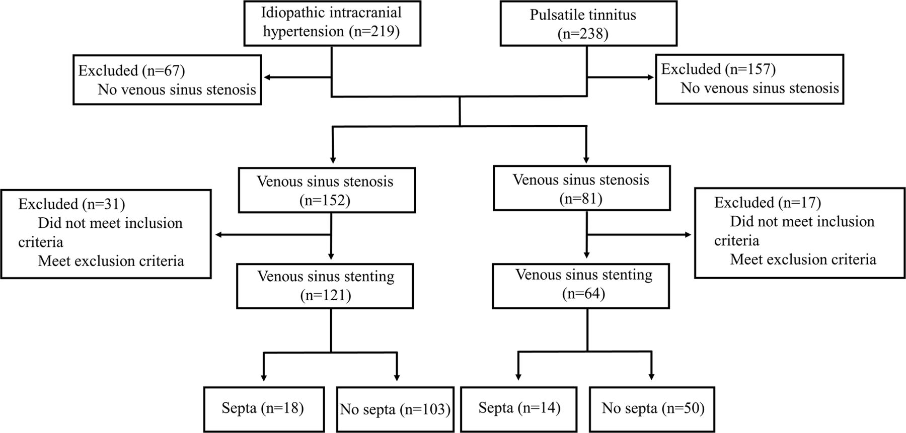
- Figure 1
Flow diagram of the study design.
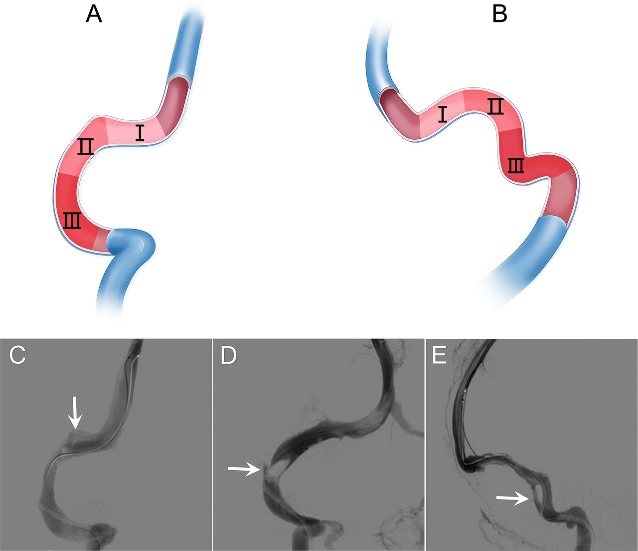
- Figure 2
The classification and DSA imaging of venous sinus septa. (A) Frontal and (B) lateral schematics of type I: septa at transverse sinus, type II: septa at transverse–sigmoid sinus junction and type III: septa at sigmoid sinus. (C) Frontal angiogram of type I. (D) Frontalangiogram of type II. (E) Lateral angiogram of type III. DSA, digital subtraction angiography.
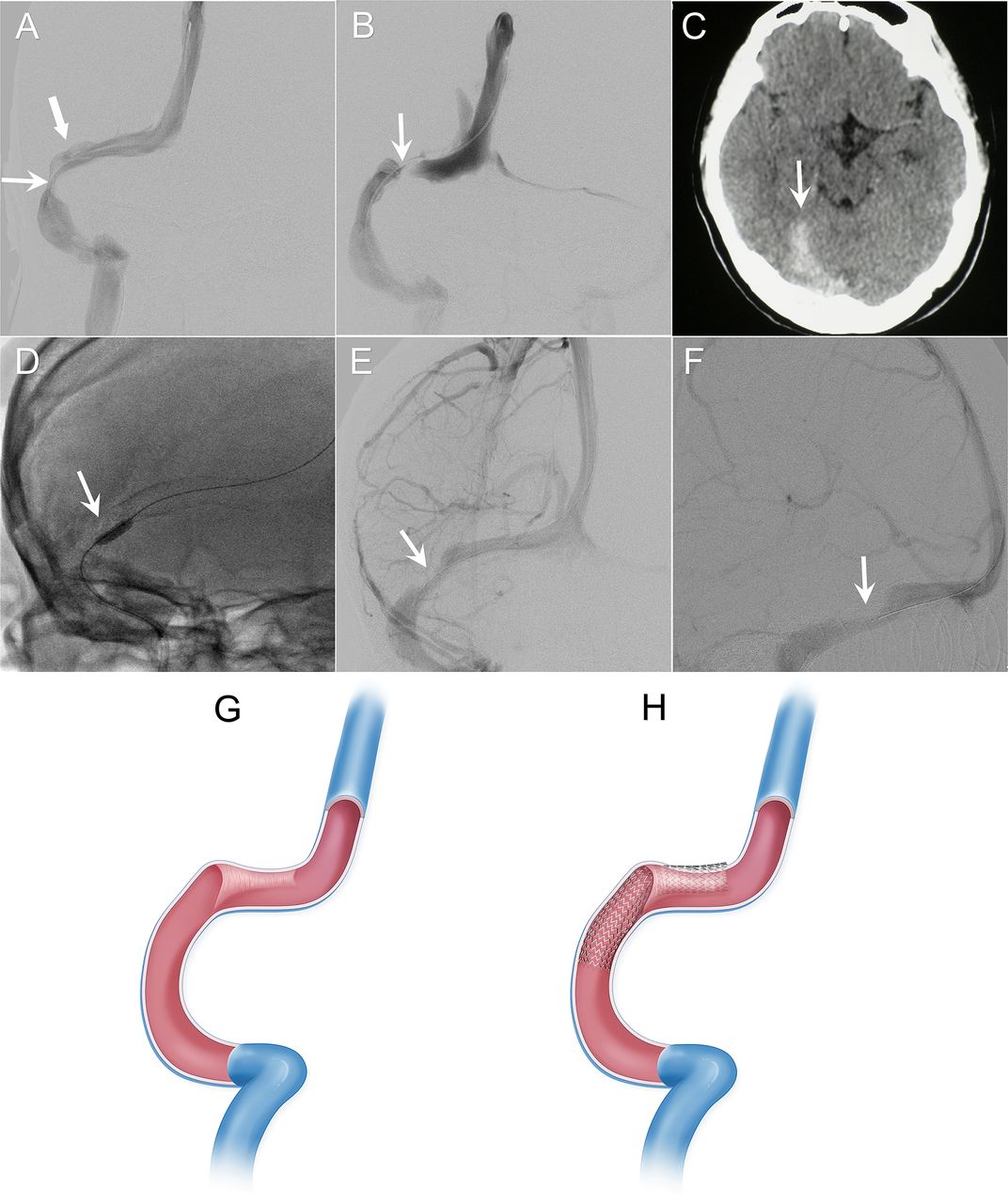
- Figure 3
Images of Case 1 and animation of complication reasoning. (A) The sinus stenosis at the right transverse–sigmoid junction (arrow) and the venous sinus septal lumen (type I, thick arrow). (B) Occluded right transverse sinus (arrow). (C) Subdural haemorrhage (arrow). (D) Dilatated balloon (arrow) in the occluded sinus segment. (E, F) Frontal and lateral angiograms of the partially recanalised venous sinus (arrow). (G) The schematic of a septum at the right transverse sinus (type I). (H) The schematic of the occluded and torn transverse sinus after stenting into the septal lumen.
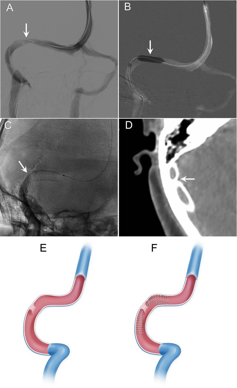
- Figure 4
Images of Case 2 and animation of complication reasoning. (A) The sinus stenosis at the right transverse–sigmoid junction (arrow). (B) Fully dilated balloon at the stenotic segment (arrow). (C) Fluoroscopy image of an incompletely expanded stent (arrow). (D) VasoCT scan image of the stent cramped at the central segment (arrow). (E) The schematic of a septum at the transverse–sigmoid sinus junction (type II). (F) The schematic of the incomplete stent expansion at the septal lumen.
- Figure 5
Images of Case 3. The sinus stenosis at the right transverse–sigmoid junction (arrow). (B) The septum at the sigmoid sinus (arrow). (C) The microcatheter through the main lumen of the sigmoid sinus (arrow). (D, E) Frontal and lateral angiograms of the expanded stent in the venous sinus (arrow). (F) Fluoroscopic image of the incompletely expanded proximal end of the stent (arrow).
- Figure 6
Animation of the slide-test technique. (A) When placed in the venous sinus lumen, the partially dilated balloon can freely slide back and forth. (B) When placed in the septal lumen, the partially dilated balloon gets stuck and should be withdrawn.


Tables
- Table 1
Baseline clinical characteristics
Clinical characteristics Means±SD (min–max) or n% Female, n (%) 30 (93.8) Age (mean±SD), years 20–57 (37.3±9. 5) Clinical duration (mean±SD), months 0.2–360 (30.0±66.2) Preoperative symptoms Headache, n (%) 10 (31.2) Dizziness, n (%) 4 (12.5) Visual dysfunction, n (%) 14 (43.8) Pulsatile tinnitus, n (%) 14 (43.8) Temporary amaurosis, n (%) 3 (9.4)


