
Main menu

  • Online first
    • Online first
  • Current issue
    • Current issue
  • Archive
    • Archive
  • Submit a paper
    • Online submission site
    • Instructions for authors
  • About the journal
    • About the journal
    • Editorial board
    • Instructions for authors
    • FAQs
    • Chinese Stroke Association
  • Help
    • Contact us
    • Feedback form
    • Reprints
    • Permissions
    • Advertising
  • BMJ Journals

User menu

  • Login

Search

  • Advanced search
  • BMJ Journals
  • Login
  • Facebook
  • Twitter
Stroke and Vascular Neurology

Advanced Search

  • Online first
    • Online first
  • Current issue
    • Current issue
  • Archive
    • Archive
  • Submit a paper
    • Online submission site
    • Instructions for authors
  • About the journal
    • About the journal
    • Editorial board
    • Instructions for authors
    • FAQs
    • Chinese Stroke Association
  • Help
    • Contact us
    • Feedback form
    • Reprints
    • Permissions
    • Advertising
Open Access

Deep white matter hyperintensity is spatially correlated to MRI-visible perivascular spaces in cerebral small vessel disease on 7 Tesla MRI

Yajing Huo, Yilin Wang, Cen Guo, Qianyun Liu, Lili Shan, Mingyuan Liu, Haibo Wu, Guanwu Li, Huihui Lv, Lingdan Lu, Yintin Zhou, Jianfeng Feng, Yan Han
DOI: 10.1136/svn-2022-001611 Published 25 April 2023
Yajing Huo
1 Department of Neurology, Yueyang Hospital of Integrated Traditional Chinese and Western Medicine, Shanghai University of Traditional Chinese Medicine, Shanghai, China
  • Find this author on Google Scholar
  • Find this author on PubMed
  • Search for this author on this site
  • ORCID record for Yajing Huo
Yilin Wang
2 Georgetown Preparatory School, North Bethesda, Maryland, USA
  • Find this author on Google Scholar
  • Find this author on PubMed
  • Search for this author on this site
Cen Guo
1 Department of Neurology, Yueyang Hospital of Integrated Traditional Chinese and Western Medicine, Shanghai University of Traditional Chinese Medicine, Shanghai, China
  • Find this author on Google Scholar
  • Find this author on PubMed
  • Search for this author on this site
  • ORCID record for Cen Guo
Qianyun Liu
1 Department of Neurology, Yueyang Hospital of Integrated Traditional Chinese and Western Medicine, Shanghai University of Traditional Chinese Medicine, Shanghai, China
  • Find this author on Google Scholar
  • Find this author on PubMed
  • Search for this author on this site
Lili Shan
1 Department of Neurology, Yueyang Hospital of Integrated Traditional Chinese and Western Medicine, Shanghai University of Traditional Chinese Medicine, Shanghai, China
  • Find this author on Google Scholar
  • Find this author on PubMed
  • Search for this author on this site
Mingyuan Liu
1 Department of Neurology, Yueyang Hospital of Integrated Traditional Chinese and Western Medicine, Shanghai University of Traditional Chinese Medicine, Shanghai, China
  • Find this author on Google Scholar
  • Find this author on PubMed
  • Search for this author on this site
Haibo Wu
1 Department of Neurology, Yueyang Hospital of Integrated Traditional Chinese and Western Medicine, Shanghai University of Traditional Chinese Medicine, Shanghai, China
  • Find this author on Google Scholar
  • Find this author on PubMed
  • Search for this author on this site
Guanwu Li
3 Department of Radiology, Yueyang Hospital of Integrated Traditional Chinese and Western Medicine, Shanghai University of Traditional Chinese Medicine, Shanghai, China
  • Find this author on Google Scholar
  • Find this author on PubMed
  • Search for this author on this site
Huihui Lv
1 Department of Neurology, Yueyang Hospital of Integrated Traditional Chinese and Western Medicine, Shanghai University of Traditional Chinese Medicine, Shanghai, China
  • Find this author on Google Scholar
  • Find this author on PubMed
  • Search for this author on this site
Lingdan Lu
1 Department of Neurology, Yueyang Hospital of Integrated Traditional Chinese and Western Medicine, Shanghai University of Traditional Chinese Medicine, Shanghai, China
  • Find this author on Google Scholar
  • Find this author on PubMed
  • Search for this author on this site
Yintin Zhou
1 Department of Neurology, Yueyang Hospital of Integrated Traditional Chinese and Western Medicine, Shanghai University of Traditional Chinese Medicine, Shanghai, China
  • Find this author on Google Scholar
  • Find this author on PubMed
  • Search for this author on this site
Jianfeng Feng
4 Institute of Science and Technology for Brain-inspired Intelligence, Fudan University, Shanghai, China
  • Find this author on Google Scholar
  • Find this author on PubMed
  • Search for this author on this site
Yan Han
1 Department of Neurology, Yueyang Hospital of Integrated Traditional Chinese and Western Medicine, Shanghai University of Traditional Chinese Medicine, Shanghai, China
  • Find this author on Google Scholar
  • Find this author on PubMed
  • Search for this author on this site
  • Article
  • Figures & Data
  • eLetters
  • Info & Metrics
  • PDF
Loading

Article Figures & Data

Figures

  • Tables
  • Figure 1
    • Download figure
    • Open in new tab
    • Download powerpoint
    Figure 1

    Flow chart of enrollment in this study. WMH, white matter hyperintensity.
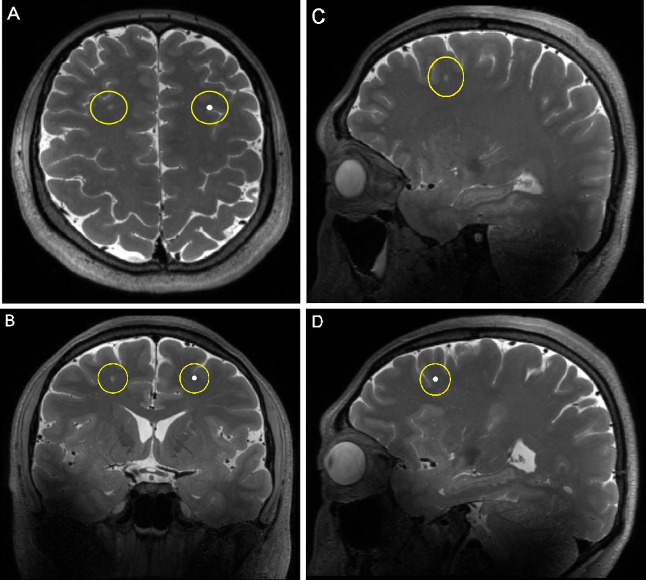
  • Figure 2
    • Download figure
    • Open in new tab
    • Download powerpoint
    Figure 2

    Illustration of reference sites of dWMH. The degrees of the dilatation and number of PVS within a spherical area with a radius of 1 cm around dWMH and the anatomically corresponding reference area in the contralateral hemisphere were evaluated in axil plane (A), coronal plane (B) and sagittal plane (C and D). dWMH, deep white matter hyperintensity; PVS, perivascular space.

  • Figure 3
    • Download figure
    • Open in new tab
    • Download powerpoint
    Figure 3

    The reference template used for the grading of PVS. From above to below, each template illustrates different extents of the dilatation of PVS. A single location was revealed in three directions (from left to right: axial, coronal, sagittal) in each row. PVS, perivascular space.
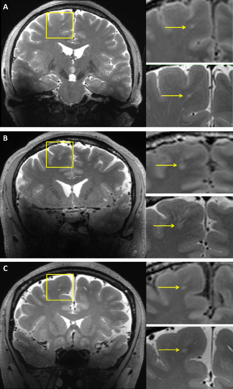
  • Figure 4
    • Download figure
    • Open in new tab
    • Download powerpoint
    Figure 4

    Illustration of spatial connections between WMH and PVS. (A) Type 1, small punctate WMH (yellow arrow) was spatially connected with one PVS tube. (B) Type 2, flake-like WMH (yellow arrow) was connected with multiple tubes. (C) Type 3, insular WMH (yellow arrow) without PVS connection. PVS, perivascular space; WMH, white matter hyperintensity.
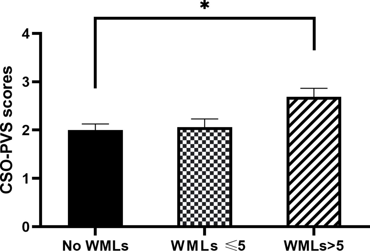
  • Figure 5
    • Download figure
    • Open in new tab
    • Download powerpoint
    Figure 5

    Comparison of CSO-PVS scores among subjects without, with ≤5 and with >5 WMLs. *indicates p<0.05. The results are showed as means±SEM. CSO-PVS, centrum semiovale-perivascular space; WMLs, white matter lesions.

Tables

  • Figures
  • Table 1

    Baseline characteristics of patients with WMHs and controls without WMHs

    CategoryPatients with WMHsControls (n=16)
    P value
    ≤5 WMLs (n=16)>5 WMLs (n=16)
    Age (mean±SD, year)44.06±6.1344.00±6.8942.75±6.260.81
    Female, n (%)7 (43.8)6 (37.5)6 (37.5)0.917
    Current smoker, n (%)3 (18.8)3 (18.8)2 (12.5)1
    Alcohol user, n (%)0 (0.0)1 (6.3)3 (18.8)0.304
    Hypertension, n (%)1 (6.3)3 (18.8)2 (12.5)0.859
    Diabetes mellitus, n (%)0 (0.0)0 (0.0)1 (6.3)1
    Hyperlipidaemia, n (%)2 (12.5)5 (31.3)3 (18.8)0.556
    Lacunes, n(%)0 (0.0)3 (18.6)0 (0.0)0.097
    CMBs, n(%)1 (6.3)1 (6.3)0 (0.0)1
    • CMBs, cerebral microbleeds; WMH, white matter hyperintensity; WML, white matter lesion.

  • Table 2

    The degree of PVS dilatation in NAWM surrounding dWMH and reference site


    N(%)
    Reference site
    Total
    012
    dWMH
     00 (0.0%)2 (100%)0 (0.0%)2
     13 (3.2%)85 (91.4%)5 (5.4%)93
     23 (4.5%)39 (59.1%)24 (36.4%)66
     30 (0.0%)1 (33.3%)2 (66.7%)3
    total612731164
    • dWMH, deep white matter hyperintensity; NAWM, normal-appearing white matter; PVS, perivascular space.

  • Table 3

    The degree of PVS number in NAWM surrounding dWMH and reference site


    N (%)
    Reference site
    Total
    012345
    dWMH
     00 (0.0%)2 (100%)0 (0.0%)0 (0.0%)0 (0.0%)0 (0.0%)2
     15 (8.9%)48 (85.7%)3 (5.4%)0 (0.0%)0 (0.0%)0 (0.0%)56
     21 (1.4%)40 (55.6%)27 (37.5%)4 (5.6%)0 (0.0%)0 (0.0%)72
     30 (0.0%)7 (30.4%)14 (60.9%)2 (8.7%)0 (0.0%)0 (0.0%)23
     40 (0.0%)2 (25.0%)2 (25.0%)4 (50.0%)0 (0.0%)0 (0.0%)8
     50 (0.0%)0 (0.0%)1 (33.3%)0 (0.0%)1 (33.3%)1 (33.3%)3
    Total699471011164
    • dWMH, deep white matter hyperintensity; NAWM, normal-appearing white matter; PVS, perivascular space.

PreviousNext
Back to top
Vol 8 Issue 2 Table of Contents
Stroke and Vascular Neurology: 8 (2)
  • Table of Contents
  • Table of Contents (PDF)
  • About the Cover
  • Index by author
  • Front Matter (PDF)
Email

Thank you for your interest in spreading the word on Stroke and Vascular Neurology.

NOTE: We only request your email address so that the person you are recommending the page to knows that you wanted them to see it, and that it is not junk mail. We do not capture any email address.

Enter multiple addresses on separate lines or separate them with commas.
Deep white matter hyperintensity is spatially correlated to MRI-visible perivascular spaces in cerebral small vessel disease on 7 Tesla MRI
(Your Name) has sent you a message from Stroke and Vascular Neurology
(Your Name) thought you would like to see the Stroke and Vascular Neurology web site.
CAPTCHA
This question is for testing whether or not you are a human visitor and to prevent automated spam submissions.
Print
Alerts
Sign In to Email Alerts with your Email Address
Citation Tools
Deep white matter hyperintensity is spatially correlated to MRI-visible perivascular spaces in cerebral small vessel disease on 7 Tesla MRI
Yajing Huo, Yilin Wang, Cen Guo, Qianyun Liu, Lili Shan, Mingyuan Liu, Haibo Wu, Guanwu Li, Huihui Lv, Lingdan Lu, Yintin Zhou, Jianfeng Feng, Yan Han
Stroke and Vascular Neurology Apr 2023, 8 (2) 144-150; DOI: 10.1136/svn-2022-001611

Citation Manager Formats

  • BibTeX
  • Bookends
  • EasyBib
  • EndNote (tagged)
  • EndNote 8 (xml)
  • Medlars
  • Mendeley
  • Papers
  • RefWorks Tagged
  • Ref Manager
  • RIS
  • Zotero
Cite This
  • APA
  • Chicago
  • Endnote
  • MLA
Loading
Deep white matter hyperintensity is spatially correlated to MRI-visible perivascular spaces in cerebral small vessel disease on 7 Tesla MRI
Yajing Huo, Yilin Wang, Cen Guo, Qianyun Liu, Lili Shan, Mingyuan Liu, Haibo Wu, Guanwu Li, Huihui Lv, Lingdan Lu, Yintin Zhou, Jianfeng Feng, Yan Han
Stroke and Vascular Neurology Apr 2023, 8 (2) 144-150; DOI: 10.1136/svn-2022-001611
Download PDF

Share
Deep white matter hyperintensity is spatially correlated to MRI-visible perivascular spaces in cerebral small vessel disease on 7 Tesla MRI
Yajing Huo, Yilin Wang, Cen Guo, Qianyun Liu, Lili Shan, Mingyuan Liu, Haibo Wu, Guanwu Li, Huihui Lv, Lingdan Lu, Yintin Zhou, Jianfeng Feng, Yan Han
Stroke and Vascular Neurology Apr 2023, 8 (2) 144-150; DOI: 10.1136/svn-2022-001611
Reddit logo Twitter logo Facebook logo Mendeley logo
Respond to this article
  • Tweet Widget
  • Facebook Like
  • Google Plus One
  • Article
    • Abstract
    • Introduction
    • Methods
    • Results
    • Discussion
    • Conclusion
    • Data availability statement
    • Ethics statements
    • Footnotes
    • References
  • Figures & Data
  • eLetters
  • Info & Metrics
  • PDF

Related Articles

Cited By...

More in this TOC Section

  • Stepwise improvement in intracerebral haematoma expansion prediction with advanced imaging: a comprehensive comparison of existing scores
  • Learning curve and embolisation strategy in single-stage surgery combined embolisation and microsurgery for brain arteriovenous malformations: results from a nationwide multicentre prospective registry study
  • Thrombus iodine-based perviousness is associated with recanalisation and functional outcomes in endovascular thrombectomy
Show more Original research

Similar Articles


CONTENT

  • Latest content
  • Current issue
  • Archive
  • eLetters
  • Sign up for email alerts
  • RSS

JOURNAL

  • About the journal
  • Editorial board
  • Recommend to librarian
  • Chinese Stroke Association

AUTHORS

  • Instructions for authors
  • Submit a paper
  • Track your article
  • Open Access at BMJ

HELP

  • Contact us
  • Reprints
  • Permissions
  • Advertising
  • Feedback form

© 2025 Chinese Stroke Association