

Article Figures & Data
Figures
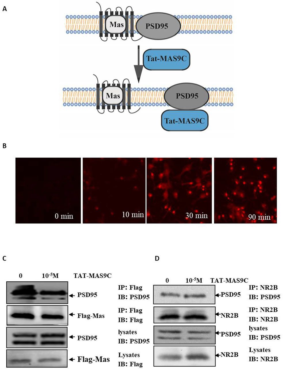
- Figure 1
TAT-MAS9C destroys the interaction between PSD95 and Mas. (A) Hypothesis: the Mas–PSD95 complex may be disrupted by TAT-MAS9C. (B) The rhodamine-labelled TAT-MAS9C (10−5 M) accumulation in neurons was visualised after incubation for 0, 10, 30 and 90 min. (C) PSD95 was subjected to coimmunoprecipitation with Flag-Mas in HEK293 cells treated with 10−5 M TAT-MAS9C. (D) PSD95 was subjected to coimmunoprecipitation with NR2B in 293 cells treated with 10−5 M TAT-MAS9C. PSD95, postsynaptic density protein-95.
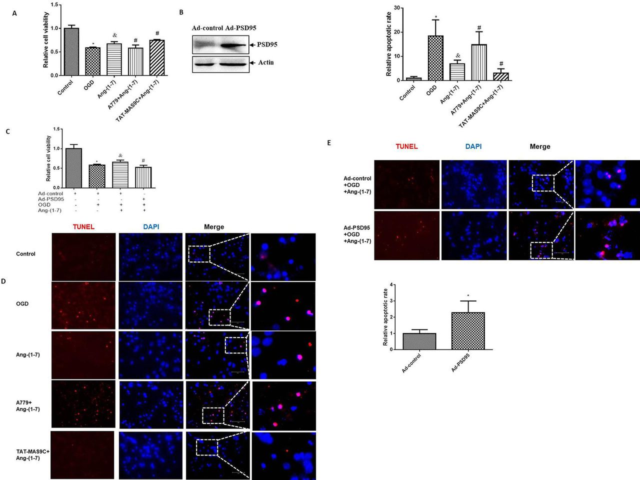
- Figure 2
Effects of PSD95 on the Ang-(1-7)–Mas-induced cerebral ischaemia protection in OGD neurons. Neurons were incubated with Ang-(1-7) (10−6 M), A779 (10−5 M) or TAT-MAS9C (10−5 M) during OGD. After OGD for 1 hour, cells were switched to normal condition for 24 hours. For Ad-PSD95 infection, neurons were infected with Ad-PSD95 or Ad-control at MOI of 100 for 3 days. Then, neurons were incubated with Ang-(1-7) (10−6 M) for 1 hour of OGD treatment and returned to normal conditions for 24 hours. (A) The effect of TAT-MAS9C on cell viability was measured using the CCK8 assay in Ang-(1-7)-treated OGD neurons. The data within each group were normalised to those of the control (n=6; vs control group*, vs OGD group&, vs Ang-(1-7) group#). (B) The overexpression of PSD95 by Ad-PSD95 infection was detected by western blot. (C) The effect of Ad-PSD95 on cell viability was measured using the CCK8 assay in Ang-(1-7)-treated OGD neurons (n=6, vs Ad-control*, vs OGD group&, vs Ad-control+Ang-(1-7)# group). (D) The effect of TAT-MAS9C on the apoptosis of neurons was identified by TUNEL staining and quantified using the percentage of TUNEL-positive cells. The data within each group were normalised to those in the control group. (E) The effect of PSD95 overexpression on the apoptosis of neurons was identified by TUNEL staining and quantified using the percentage of TUNEL-positive cells. The data within each group were normalised to those in the Ad-control group. *P<0.05, &P<0.05, #P<0.05. Ad-PSD95, PSD95 adenovirus; Ang-(1-7), angiotensin-(1-7); OGD, oxygen–glucose deprivation; PSD95, postsynaptic density protein-95; TUNEL, terminal deoxynucleotidyl transferase-mediated dUTP nick-end labelling; DAPI, 4',6-diamidino-2-phenylindole.
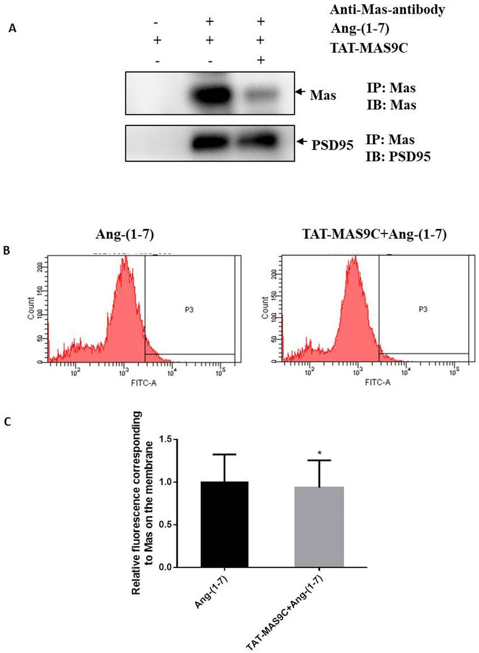
- Figure 3
Effect of TAT-MAS9C on the cell membrane localisation of Mas protein in OGD neurons treated with Ang-(1-7). Neurons or NE-4C cells were treated with OGD for 1 hour and then stimulated for 25 min with 10−6 M Ang-(1-7) or 10−5 M TAT-MAS9C+10−6 M Ang-(1-7) in HBSS. After washing with cold HBSS, cells were treated with anti-Mas antibody for 50 min on ice. (A) Protein immunoprecipitation was used to detect the expression level of MAS in the plasma membrane of neurons. Cell lysates were subjected to immunoprecipitation with protein A/G. The expression levels of MAS and PSD95 in the immunoprecipitation complex was detected by western blot. Images were representative of three independent experiments. (B) Flow cytometry was used to detect the expression level of MAS in the plasma membrane of NE-4C cells. Cells were treated with Alexa Fluor 488 goat anti-rabbit IgG for 50 min on ice, washed with cold HBSS, and digested with 5 mM EDTA in PBS on ice. Suspended NE-4C cells were collected and detected by flow cytometry. (C) The relative fluorescence corresponding to MAS on the membrane was analysed by flow cytometry. Values were obtained from three separate experiments (n=3). Ang-(1-7), angiotensin-(1-7); IB, immunoblot; IP, immunoprecipitation; OGD, oxygen–glucose deprivation; PSD95, postsynaptic density protein-95.
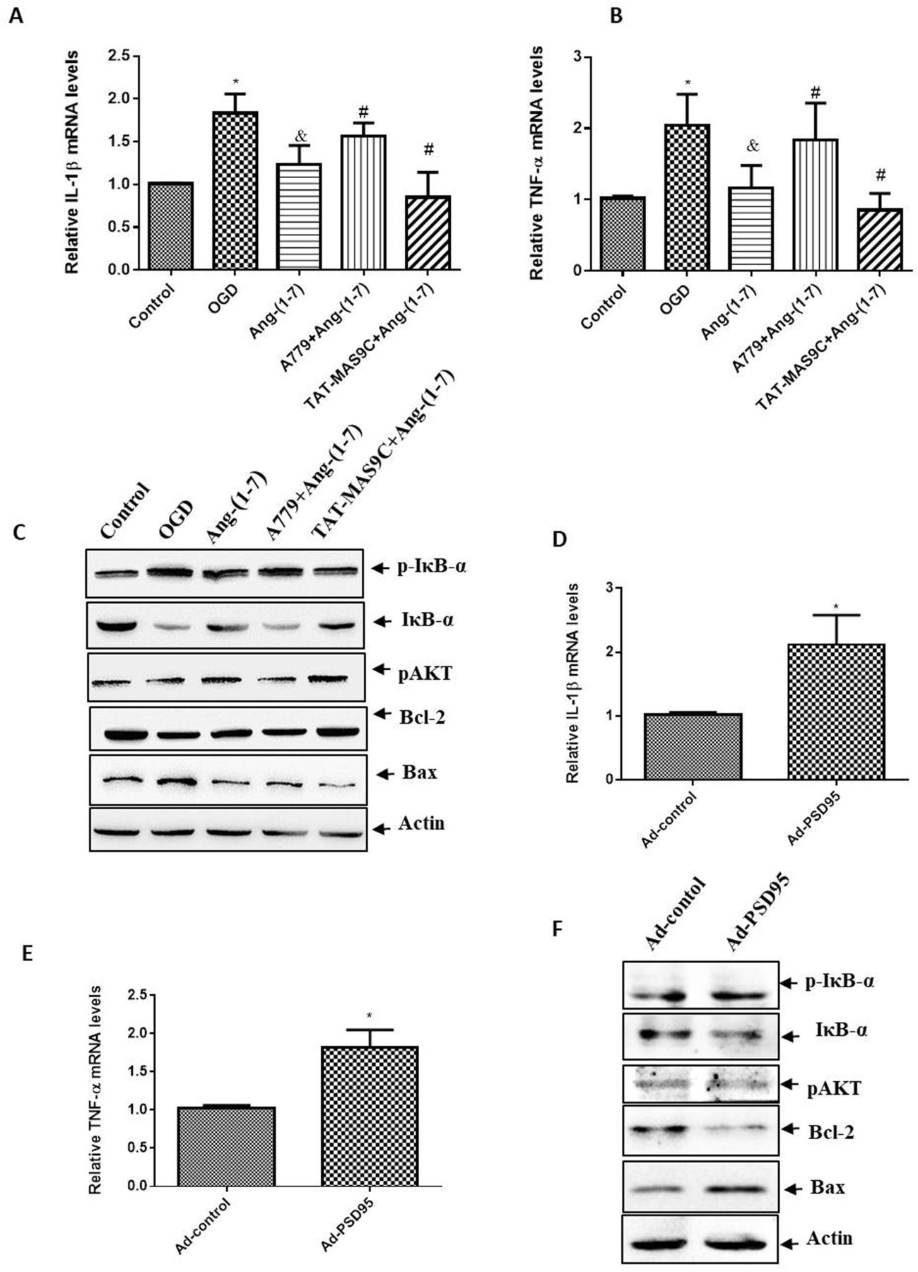
- Figure 4
Effect of PSD95 on the signalling pathway in Ang-(1-7)-treated neurons. Neurons were incubated with Ang-(1-7) (10−6 M), A779 (10−5 M) or TAT-MAS9C (10−5 M) during OGD. After OGD for 1 hour, cells were switched to normal conditions for 24 hours. qPCR was used to analyse the expression levels of (A) IL-1β and (B) TNF-α. Values were expressed as mean±SD (n=3; vs control group*, vs OGD group&, vs Ang-(1-7) group#). (C) Western blot was used to analyse the expression levels of p-IκB-α, IκB-α, p-AKT, Bcl-2 and Bax. Images were representative of three independent experiments. For Ad-PSD95 infection, neurons were infected with Ad-PSD95 or Ad-control at MOI of 100 for 3 days. Then, neurons were incubated with Ang-(1-7) (10−6 M) for 1 hour of OGD treatment and returned to normal conditions for 24 hours. qPCR was used to analyse the expression levels of (D) IL-1β and (E) TNF-α. Values were expressed as mean±SD (n=3, vs Ad-control*). (F) Western blot was used to analyse the expression levels of p-IκB-α, IκB-α, p-AKT, Bcl-2 and Bax. Images were representative of three independent experiments. *P<0.05, &P<0.05, #P<0.05. Ad-PSD95, PSD95 adenovirus; Ang-(1-7), angiotensin-(1-7); IL, interleukin; OGD, oxygen–glucose deprivation; PSD95, postsynaptic density protein-95; qPCR, quantitative PCR; TNF-α, tumour necrosis factor alpha.
- Figure 5
Effects of PSD95 on Ang-(1-7)–Mas-induced cerebral ischaemia protection in MCAO rats. Rats were pretreated with artificial cerebrospinal fluid, Ang-(1-7) or Ang-(1-7)+TAT-MAS9C for 7 days prior to MCAO. Rats were then subjected to MCAO for 90 min. Ang-(1-7), A779 or TAT-MAS9C was injected slowly into the lateral ventricle 15 min after MCAO once a day. (A) The neurological performance of rats was graded using Longa’s test 30 hours after MCAO. (B) The infarct volume was evaluated 30 hours after MCAO. Brain tissue sections were stained with TTC. (C) The infarct volume was calculated as the ratio of the infarct area to the total area by using ImageJ software. (D) Western blot was used to analyse the expression levels of Bcl-2, Bax and cleaved caspase 3 in the ischaemic penumbra of the cerebral cortex. (E)The Morris water maze test was performed to assess spatial memory. Representative images are swimming tracks. (F) The number of crossings over the platform area in the Morris water maze test was analysed. Values were expressed as mean±SD (n=6, vs sham group*, vs MCAO group&, vs Ang-(1-7) group#). *P<0.05, &P<0.05, #P<0.05. Ang-(1-7), angiotensin-(1-7); MCAO, middle cerebral artery occlusion; PSD95, postsynaptic density protein-95; TTC, 2,3,5-triphenyltetrazolium chloride.


