
Main menu

  • Online first
    • Online first
  • Current issue
    • Current issue
  • Archive
    • Archive
  • Submit a paper
    • Online submission site
    • Instructions for authors
  • About the journal
    • About the journal
    • Editorial board
    • Instructions for authors
    • FAQs
    • Chinese Stroke Association
  • Help
    • Contact us
    • Feedback form
    • Reprints
    • Permissions
    • Advertising
  • BMJ Journals

User menu

  • Login

Search

  • Advanced search
  • BMJ Journals
  • Login
  • Facebook
  • Twitter
Stroke and Vascular Neurology

Advanced Search

  • Online first
    • Online first
  • Current issue
    • Current issue
  • Archive
    • Archive
  • Submit a paper
    • Online submission site
    • Instructions for authors
  • About the journal
    • About the journal
    • Editorial board
    • Instructions for authors
    • FAQs
    • Chinese Stroke Association
  • Help
    • Contact us
    • Feedback form
    • Reprints
    • Permissions
    • Advertising
Open Access

Unifying theory of carotid plaque disruption based on structural phenotypes and forces expressed at the lumen/wall interface

Luis Savastano, Hossein Mousavi, Yang Liu, Siri Sahib S Khalsa, Yihao Zheng, Evan Davis, Adithya Reddy, Waleed Brinjikji, Ankur Bhambri, Joshua Cockrum, Aditya S Pandey, B Gregory Thompson, David Gordon, Eric J Seibel, Howard Yonas
DOI: 10.1136/svn-2021-001451 Published 20 December 2022
Luis Savastano
1 Neurosurgery, Mayo Clinic, Rochester, Minnesota, USA
  • Find this author on Google Scholar
  • Find this author on PubMed
  • Search for this author on this site
Hossein Mousavi
2 Department of Neurology, University of New Mexico School of Medicine, Albuquerque, New Mexico, USA
  • Find this author on Google Scholar
  • Find this author on PubMed
  • Search for this author on this site
Yang Liu
3 Radiology, Mayo Clinic, Rochester, Minnesota, USA
  • Find this author on Google Scholar
  • Find this author on PubMed
  • Search for this author on this site
  • ORCID record for Yang Liu
Siri Sahib S Khalsa
4 Department of Neurosurgery, University of Michigan, Ann Arbor, Michigan, USA
  • Find this author on Google Scholar
  • Find this author on PubMed
  • Search for this author on this site
Yihao Zheng
5 Department of Mechanical Engineering, Worcester Polytechnic Institute, Worcester, Massachusetts, USA
  • Find this author on Google Scholar
  • Find this author on PubMed
  • Search for this author on this site
  • ORCID record for Yihao Zheng
Evan Davis
4 Department of Neurosurgery, University of Michigan, Ann Arbor, Michigan, USA
  • Find this author on Google Scholar
  • Find this author on PubMed
  • Search for this author on this site
Adithya Reddy
4 Department of Neurosurgery, University of Michigan, Ann Arbor, Michigan, USA
  • Find this author on Google Scholar
  • Find this author on PubMed
  • Search for this author on this site
Waleed Brinjikji
6 Department of Radiology, Mayo Clinic, Rochester, Minnesota, USA
  • Find this author on Google Scholar
  • Find this author on PubMed
  • Search for this author on this site
Ankur Bhambri
4 Department of Neurosurgery, University of Michigan, Ann Arbor, Michigan, USA
  • Find this author on Google Scholar
  • Find this author on PubMed
  • Search for this author on this site
Joshua Cockrum
4 Department of Neurosurgery, University of Michigan, Ann Arbor, Michigan, USA
  • Find this author on Google Scholar
  • Find this author on PubMed
  • Search for this author on this site
Aditya S Pandey
7 Neurosurgery, Michigan Medicine, Ann Arbor, Michigan, USA
8 Radiology, Michigan Medicine, Ann Arbor, Michigan, USA
  • Find this author on Google Scholar
  • Find this author on PubMed
  • Search for this author on this site
B Gregory Thompson
4 Department of Neurosurgery, University of Michigan, Ann Arbor, Michigan, USA
  • Find this author on Google Scholar
  • Find this author on PubMed
  • Search for this author on this site
David Gordon
4 Department of Neurosurgery, University of Michigan, Ann Arbor, Michigan, USA
  • Find this author on Google Scholar
  • Find this author on PubMed
  • Search for this author on this site
Eric J Seibel
9 Department of Mechanical Engineering, University of Washington, Seattle, Washington, USA
  • Find this author on Google Scholar
  • Find this author on PubMed
  • Search for this author on this site
Howard Yonas
2 Department of Neurology, University of New Mexico School of Medicine, Albuquerque, New Mexico, USA
  • Find this author on Google Scholar
  • Find this author on PubMed
  • Search for this author on this site
  • Article
  • Figures & Data
  • eLetters
  • Info & Metrics
  • PDF
Loading

Article Figures & Data

Figures

  • Tables
  • Supplementary Materials
  • Additional Files
  • Figure 1
    • Download figure
    • Open in new tab
    • Download powerpoint
    Figure 1

    Phenotypes of carotid endarterectomy specimens. Preoperative computational tomographic angiography in axial (A) and sagittal reconstructions (B) reveal mixed density atheroma (yellow asterisk) leading to >70% stenosis in the common carotid artery (CCA) and proximal internal carotid artery (ICA). At surgery, the CCA, external and carotid arteries (ECA and ICA) are exposed and atheromas removed en bloc without luminal violation and minimising disruption (D). The lumen/wall interface of each specimens were imaged with an operative microscope (×25 magnification) from the flow inlet (E) and outlet (F) perspective. Based on the findings and the preoperative images, plaques were dissected both longitudinally (G) and/or transversally to optimally visualise the interface between the lumen and the atheroma (yellow asterisk). In the cohort, four recurrent phenotypes were identified: (H) Atheroma with no discontinuity of the fibrous cap; (I) atheroma with ulcer (ie, loss of continuity of the fibrous cap, generally round shaped or oval) upstream (92%), at (6%) or downstream (1%) to the point of maximal stenosis (asterisk); (J) atheroma with an intraparenchymal haemorrhage, in continuity with an ulceration in 92% of cases; (K) atheroma with false luminal formation (excavation within the plaque). Atheroma subcomponent in (H)–(K) are color coded: white for fibrous cap, light blue for calcified tissue, grey for necrotic core and red for thrombus.

  • Figure 2
    • Download figure
    • Open in new tab
    • Download powerpoint
    Figure 2

    Morphological analysis of plaque phenotypes contextualised to individualised haemodynamic conditions. Plaque ulcerations (A1) were identified as full thickness discontinuity of the fibrous cap of the atheroma per angioscopy (A2) and remained occult to CTA (arrowhead in (A3) points the approximate location of the ulcer identified per angioscopy in (A2)). CFD simulations revealed a region of local high dynamic pressure directed into the plaque in the same region of the ulcer ((A4) and arrowhead in insert). Blind-ended false luminal formation (FLF) (B1) was identified by SFE angioscopy as a crater excavating the atheroma from an ulcerative region generally associated with haematomas (arrowhead in (B2)). These excavations in the atheroma corresponded to deep surface irregularities in CTA (arrowhead in (B3)). CFD simulation in FLF demonstrated a region of local high dynamic pressure directed cranially ((B4) and arrowhead in insert). In shallow excavations, the jet of blood chisels the plaque core with high dynamic pressure (B5) and following a vigorous stream vortex (B6). In deeper excavations, the dynamic pressure of the blood significantly tapers off toward the apex of the crater (arrowhead in (B7)) and the jet of blood becomes slow and disorganised with stagnation towards the apex of the excavation (B8). Fenestrated FLF (C1) was identified per angioscopy as deep and broad excavations, usually parallel to the true lumen, with one or more opening communicating the false with the true lumen (arrowheads in (C2)). False lumens running through atheroma were also identified in CTA (inlet and outlet with arrowheads, point of maximal stenosis with asterisk in (C3)). In these cases, CFD simulation revealed localised regions of high dynamic pressure within the plaque’s FLF with maximal stress over the remaining fibrous cap from both the luminal and pseudoluminal surfaces (yellow and black arrowheads, respectively, in (C4)). Plaque haematomas (D1) were generally exposed at the lumen/wall interface and their geographic distribution and thrombus type readily identified by angioscopy (D2). Plaque haematomas frequently had an intraluminal extension with cranial propagation into the internal carotid artery (ICA) (arrowhead in a CTA in (D3)). In cases of critical stenosis, CFD simulation revealed a region of high dynamic pressure at the POMS with propagation to one side of the arterial wall circumference and minimal haemodynamic stress in the opposite side (arrowheads point high stress at the POMS and downstream in (D4)). As the flow passes through the narrowed luminal corridor, it is directed towards one side of the arterial lumen leaving the opposite side in relative stagnation, which favours clot formation (arrowheads in (D5)). Atheroma subcomponent in (A1–D1) are color coded: white for fibrous cap, light blue for calcified tissue, grey for necrotic core and red for thrombus. Refer to the online supplemental videos for the dynamic colour bars of the contour map of the CFD. ECA, external carotid artery.
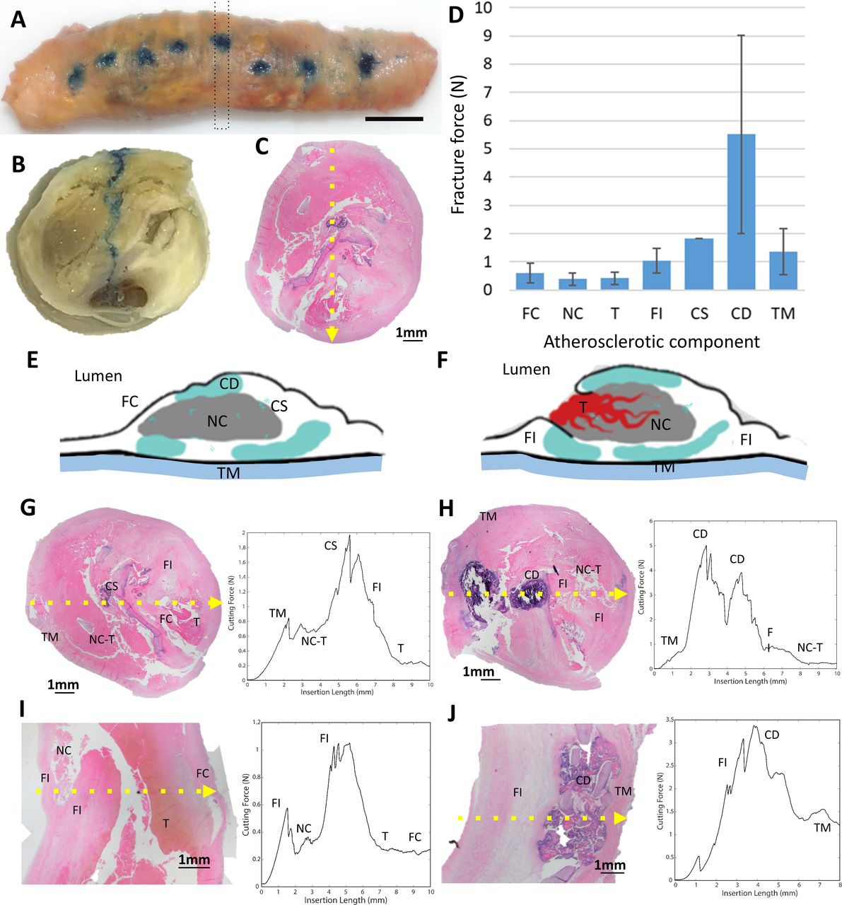
  • Figure 3
    • Download figure
    • Open in new tab
    • Download powerpoint
    Figure 3

    Mechanical testing of atherosclerotic components of carotid plaques: needle penetration tests of atheroma was conducted in a custom-made platform (blue dots in (A) corresponds to insertion points in atheroma). The position of the needle and force data were recorded at a frequency of at 50 Hz and then datasets correlated with the corresponding atheroma cross section (dotted rectangle in (A) and cross section in (B)). Tissue correlation was refined by histological analysis of the atheroma section (note: blue trajectory of needle in (B) and corresponding histological cut in (C)). The number of cutting force datapoints, the mean cutting force and SD among the datapoints for each arterial and atherosclerotic component were calculated (D). The tissues analysed in the cohort (E–F) were: fibrous cap (FC), necrotic core (NC), thrombus (T), fibrotic intima (FI), calcification speckled (CS), calcification dense (CD) and tunica media (TM). Cutting force curves with histological correlation of representative advanced and complicated carotid atherosclerosis (complex atherothrombotic lesion in (G) and partially calcified in (H), thromboatheroma in (I) and calcified fibroatheroma in (J)) are presented (dotted yellow arrow in (C) and (G–J) indicates pathway of the needle).
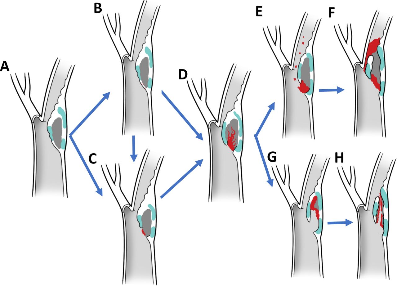
  • Figure 4
    • Download figure
    • Open in new tab
    • Download powerpoint
    Figure 4

    Biomechanical paradigm of plaque disruption with phenotypic hallmarks and driving forces. In this paradigm, plaque disruption starts on a thin-walled atheroma (A) with a fissure (B) or ulceration of the fibrous cap (C), followed by excavation of the necrotic core with formation of a plaque haematoma (D). If pro-occlusive conditions prevail, thrombosis progresses leading to luminal extension of the thrombus, narrowing and occlusion (E,F). If proexcavatory conditions prevail, the necrotic core is progressively excavated and plaque content embolised downstream mixed with thrombi resulting in blind-ended or double-ended false luminal formations (G,H).

Tables

  • Figures
  • Supplementary Materials
  • Additional Files
  • Table 1

    Structural analysis of a cohort of carotid endarterectomy specimens

    Plaque phenotypeDescriptive architectureTotal (percentage)
    Ulceration
    283 (82%)
    Relationship to point of maximal stenosis (POMS)Upstream to POMS261 (92%)
    At the POMS17 (6%)
    Downstream to POMS3 (1%)
    Upstream and at the POMS2 (1%)
    Necrotic core exposed to the blood streamPresent161 (57%)
    Absent122 (43%)
    Intraplaque haemorrhage
    233 (68%)
    Coexistence of ulcerationYes231 (99%)
    No2 (1%)
    Relationship to lumenContained89 (38%)
    Exposed144 (62%)
    Relationship to ulcerationContiguous215 (92%)
    Apart18 (8%)
    N/A6
    Relationship of haemorrhage to the downstream ending of a plaqueNot reaching159 (68%)
    Reaching65 (28%)
    Extruding beyond the plaque ending into the lumen9 (4%)
    False lumen
    165 (48%)
    Associated with thrombusYes130 (79%)
    No35 (21%)
    TypeBlind ended130 (79%)
    Fenestrated35 (21%)
  • Table 2

    Correlative analysis of plaques phenotypes with preoperative symptoms

    PatientsAsymptomaticSymptomatic
    Group A1715 (88%)2 (12%)
    Group B148 (57%)6 (43%)
    Group C259 (36%)16 (64%)
    Group D516 (12%)45 (88%)
    Total1073869
    • Group A: plaques with no disruption of the fibrous cap. Group B: plaques with ulceration. Group C: plaques with ulceration and IPH. Group D: plaques with false luminal formation.

  • Table 3

    Cutting force of individual atherosclerotic component from the needle insertion test

    ComponentNumber of datasets (penetration)Mean force (N)Std (N)
    Fibrous cap30.600.36
    Necrotic core40.390.23
    Thrombus50.420.23
    Fibrotic intima101.050.43
    Calcification speckled11.82
    Calcification dense115.523.51
    Tunica media101.350.82

Supplementary Materials

  • Figures
  • Tables
  • Additional Files
  • Supplementary data

    [svn-2021-001451supp016.pdf]

  • Supplementary video

    [svn-2021-001451supp001.mp4]

  • Supplementary video

    [svn-2021-001451supp002.mp4]

  • Supplementary video

    [svn-2021-001451supp003.mp4]

  • Supplementary video

    [svn-2021-001451supp004.mp4]

  • Supplementary video

    [svn-2021-001451supp005.mp4]

  • Supplementary video

    [svn-2021-001451supp006.mp4]

  • Supplementary video

    [svn-2021-001451supp007.mp4]

  • Supplementary video

    [svn-2021-001451supp008.mp4]

  • Supplementary video

    [svn-2021-001451supp009.mp4]

  • Supplementary video

    [svn-2021-001451supp010.mp4]

  • Supplementary video

    [svn-2021-001451supp011.mp4]

  • Supplementary video

    [svn-2021-001451supp012.mp4]

  • Supplementary video

    [svn-2021-001451supp013.mp4]

  • Supplementary video

    [svn-2021-001451supp014.mp4]

  • Supplementary video

    [svn-2021-001451supp015.mp4]

Additional Files

  • Figures
  • Tables
  • Supplementary Materials
  • Supplementary Data

    This web only file has been produced by the BMJ Publishing Group from an electronic file supplied by the author(s) and has not been edited for content.

    • Data supplement 1
    • Data supplement 2
    • Data supplement 3
    • Data supplement 4
    • Data supplement 5
    • Data supplement 6
    • Data supplement 7
    • Data supplement 8
    • Data supplement 9
    • Data supplement 10
    • Data supplement 11
    • Data supplement 12
    • Data supplement 13
    • Data supplement 14
    • Data supplement 15
    • Data supplement 16
PreviousNext
Back to top
Vol 7 Issue 6 Table of Contents
Stroke and Vascular Neurology: 7 (6)
  • Table of Contents
  • Table of Contents (PDF)
  • About the Cover
  • Index by author
  • Front Matter (PDF)
Email

Thank you for your interest in spreading the word on Stroke and Vascular Neurology.

NOTE: We only request your email address so that the person you are recommending the page to knows that you wanted them to see it, and that it is not junk mail. We do not capture any email address.

Enter multiple addresses on separate lines or separate them with commas.
Unifying theory of carotid plaque disruption based on structural phenotypes and forces expressed at the lumen/wall interface
(Your Name) has sent you a message from Stroke and Vascular Neurology
(Your Name) thought you would like to see the Stroke and Vascular Neurology web site.
CAPTCHA
This question is for testing whether or not you are a human visitor and to prevent automated spam submissions.
Print
Alerts
Sign In to Email Alerts with your Email Address
Citation Tools
Unifying theory of carotid plaque disruption based on structural phenotypes and forces expressed at the lumen/wall interface
Luis Savastano, Hossein Mousavi, Yang Liu, Siri Sahib S Khalsa, Yihao Zheng, Evan Davis, Adithya Reddy, Waleed Brinjikji, Ankur Bhambri, Joshua Cockrum, Aditya S Pandey, B Gregory Thompson, David Gordon, Eric J Seibel, Howard Yonas
Stroke and Vascular Neurology Dec 2022, 7 (6) 465-475; DOI: 10.1136/svn-2021-001451

Citation Manager Formats

  • BibTeX
  • Bookends
  • EasyBib
  • EndNote (tagged)
  • EndNote 8 (xml)
  • Medlars
  • Mendeley
  • Papers
  • RefWorks Tagged
  • Ref Manager
  • RIS
  • Zotero
Cite This
  • APA
  • Chicago
  • Endnote
  • MLA
Loading
Unifying theory of carotid plaque disruption based on structural phenotypes and forces expressed at the lumen/wall interface
Luis Savastano, Hossein Mousavi, Yang Liu, Siri Sahib S Khalsa, Yihao Zheng, Evan Davis, Adithya Reddy, Waleed Brinjikji, Ankur Bhambri, Joshua Cockrum, Aditya S Pandey, B Gregory Thompson, David Gordon, Eric J Seibel, Howard Yonas
Stroke and Vascular Neurology Dec 2022, 7 (6) 465-475; DOI: 10.1136/svn-2021-001451
Download PDF

Share
Unifying theory of carotid plaque disruption based on structural phenotypes and forces expressed at the lumen/wall interface
Luis Savastano, Hossein Mousavi, Yang Liu, Siri Sahib S Khalsa, Yihao Zheng, Evan Davis, Adithya Reddy, Waleed Brinjikji, Ankur Bhambri, Joshua Cockrum, Aditya S Pandey, B Gregory Thompson, David Gordon, Eric J Seibel, Howard Yonas
Stroke and Vascular Neurology Dec 2022, 7 (6) 465-475; DOI: 10.1136/svn-2021-001451
Reddit logo Twitter logo Facebook logo Mendeley logo
Respond to this article
  • Tweet Widget
  • Facebook Like
  • Google Plus One
  • Article
    • Abstract
    • Introduction
    • Materials and methods
    • Results
    • Discussion
    • Conclusions
    • Data availability statement
    • Ethics statements
    • Acknowledgments
    • Footnotes
    • References
  • Figures & Data
  • eLetters
  • Info & Metrics
  • PDF

Related Articles

Cited By...

More in this TOC Section

  • Learning curve and embolisation strategy in single-stage surgery combined embolisation and microsurgery for brain arteriovenous malformations: results from a nationwide multicentre prospective registry study
  • Stepwise improvement in intracerebral haematoma expansion prediction with advanced imaging: a comprehensive comparison of existing scores
  • Thrombus iodine-based perviousness is associated with recanalisation and functional outcomes in endovascular thrombectomy
Show more Original research

Similar Articles


CONTENT

  • Latest content
  • Current issue
  • Archive
  • eLetters
  • Sign up for email alerts
  • RSS

JOURNAL

  • About the journal
  • Editorial board
  • Recommend to librarian
  • Chinese Stroke Association

AUTHORS

  • Instructions for authors
  • Submit a paper
  • Track your article
  • Open Access at BMJ

HELP

  • Contact us
  • Reprints
  • Permissions
  • Advertising
  • Feedback form

© 2025 Chinese Stroke Association