

Article Figures & Data
Figures
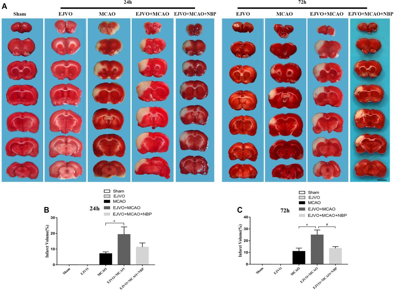
- Figure 1
Cerebral venous circulation disturbance increased infarction volume, and NBP could reduce infarct area after 3 days of treatment. (A) Infarction volume was evaluated by TTC staining 24 hours and 72 hours postoperation in groups of MCAO, EJVO+MCAO and EJVO+MCAO+ NBP. (B) Quantification of infarction volume at 24 hours after operation. (C) Quantification of infarction volume at 72 hours after operation. Data are presented as mean±SD; n=5 per group. *P<0.05, EJVO+MCAO vs MCAO; #P<0.05, EJVO+MCAO + NBP vs EJVO+MCAO. Scale bar=5 mm. EJVO, external jugular vein occlusion; MACO, middle cerebral artery occlusion; NBP, Dl-3-n-butylphthalide; TTC, 2, 3, 5-triphenyltetrazolium chloride.
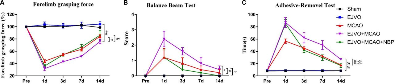
- Figure 2
Cerebral venous circulation disturbance aggravate deterioration of neurological function, and NBP treatment contributed to improved neurological function in rats of MCAO accompanied by EJVO. (A) Quantification analysis of forelimb grasping force by repeated measurement ANOVA. (B) Quantification analysis of balance beam test score by repeated measurement ANOVA. (C) Quantification analysis of balance beam test score by repeated measurement ANOVA. Data are presented as mean±SD; n=5 per group. *P<0.05, EJVO +MCAO vs sham; **P<0.01, EJVO +MCAO vs sham; #p<0.05, EJVO +MCAO vs MCAO; ##p<0.01, EJVO +MCAO vs MCAO; §P<0.05, EJVO +MCAO + NBP vs EJVO +MCAO; §§p<0.01, EJVO +MCAO + NBP vs EJVO +MCAO. ANOVA, analysis of variance; EJVO, external jugular vein occlusion; MACO, middle cerebral artery occlusion; NBP, Dl-3-n-butylphthalide.
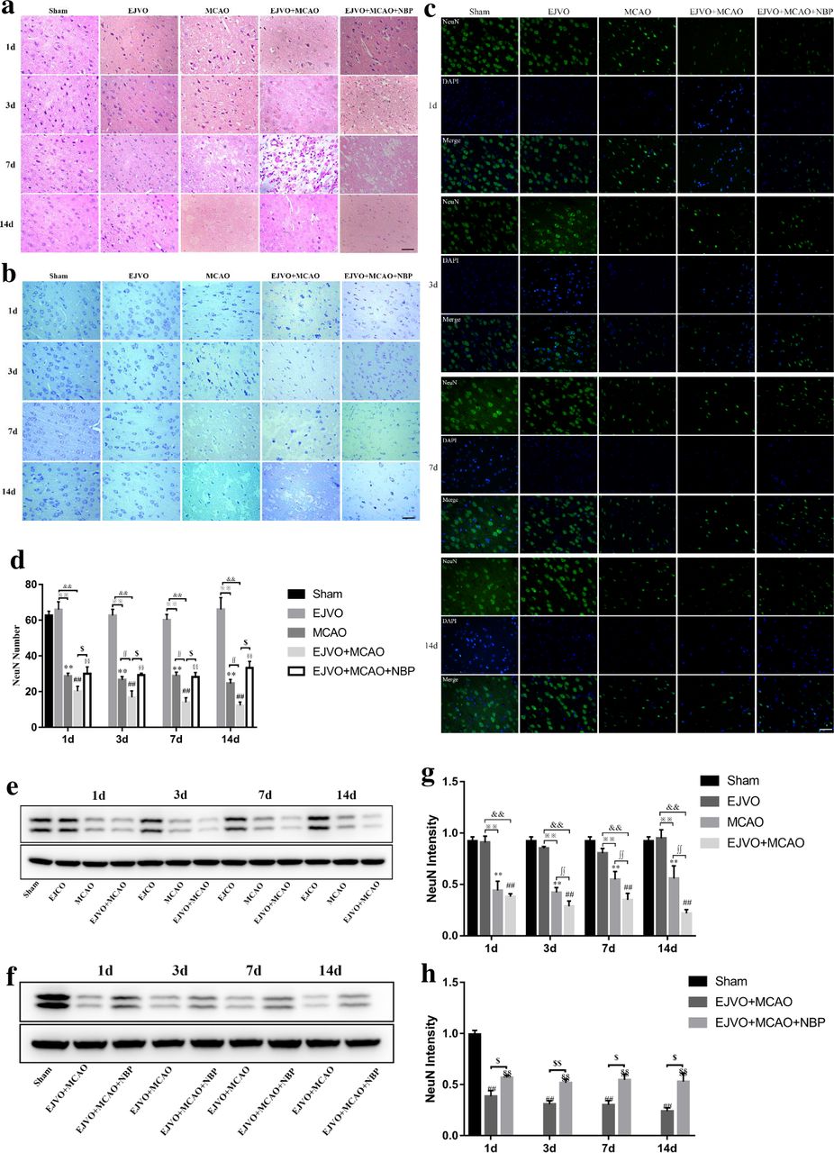
- Figure 3
Disturbance of venous circulation aggravates neuron injury after MCAO, and NBP played a neuroprotective role in rats of MCAO accompanied by EJVO. (A) Representative H&E staining showing neuron cells from first to 14th day in infarction area or corresponding area in the sham, EJVO, MCAO, EJVO +MCAO and EJVO +MCAO+ NBP groups. (B) Representative Nissl staining showing neuron cells from first to 14th day in infarction area or corresponding area in the sham, EJVO, MCAO, EJVO +MCAO and EJVO +MCAO+ NBP groups. (C) Neuron cells were labelled with NeuN antibody (green) and merged with DAPI (blue) in ischaemic area or corresponding regions of sham or EJVO group. (D) Changes of neuron cells as measured by NeuN positive cell in the different time points after operation with immunofluorescence staining. (E, F) Western blots showed NueN expression after operation treated with NBP or not. (G, H) Quantification analysis of relative expression of NeuN level. Data are presented as mean±SD, n=5 per group for immunofluorescence staining and n=3 per group for Western bolt. **P<0.01, MCAO vs Sham; ##p<0.01, EJVO +MCAO vs sham; §§p<0.01, EJVO +MCAO + NBP vs sham; ※※p<0.01, MCAO vs EJVO; &&p<0.01, EJVO +MCAO vs EJVO; ∫∫p<0.05, EJVO +MCAO vs MCAO; $p<0.05, EJVO +MCAO + NBP vs EJVO +MCAO; $$p<0.01, EJVO +MCAO + NBP vs EJVO +MCAO. Scale bar=50 µm. EJVO, external jugular vein occlusion; MACO, middle cerebral artery occlusion; NBP, Dl-3-n-butylphthalide.
- Figure 4
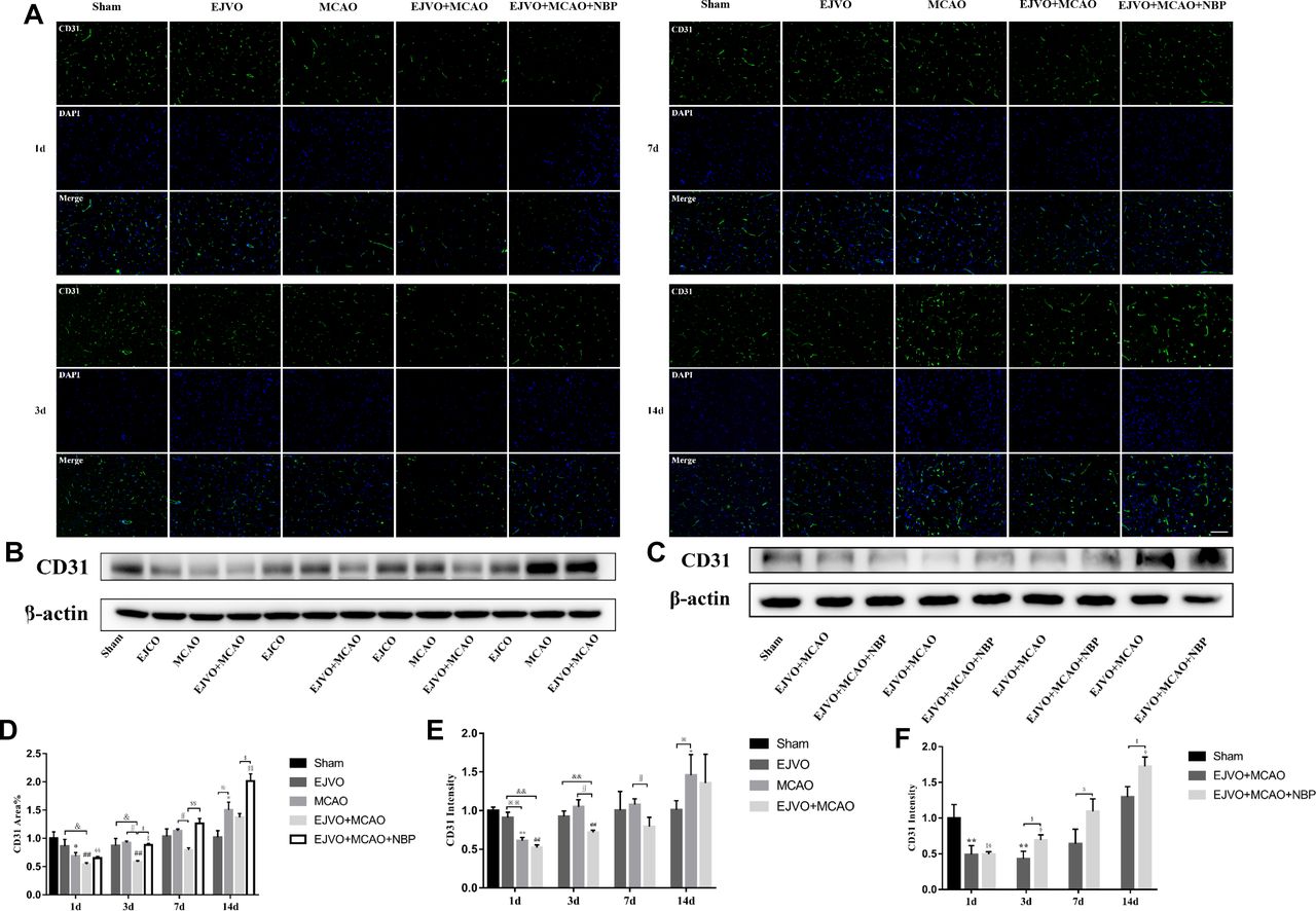
Disturbance of venous circulation decrease microvascular angiogenesis, and NBP treatment could improve angiogenesis. (A) Immunofluorescence staining of CD31 positive cells in the microvessels in the region of ischaemia from first to 14th day after operation in the sham, EJVO, MCAO, EJVO +MCAO and EJVO +MCAO+ NBP groups. (B, C) Western blots of expression of CD31. (D) Quantification of microvassels density measured by immunofluorescence staining of CD31. (E, F) Quantification analysis of relative expression of CD31 level measured by Western blot. Data are presented as mean±SD, n=5 per group for immunofluorescence staining and n=3 per group for Western bolt. *P<0.05, MCAO vs sham; **P<0.01, MCAO vs Sham; ## P<0.01, EJVO +MCAO vs sham; §p<0.05, EJVO +MCAO + NBP vs sham; §§p<0.01, EJVO +MCAO + NBP vs sham; ※P<0.05, MCAO vs EJVO; ※※P<0.01, MCAO vs EJVO; & p<0.05, EJVO +MCAO vs EJVO; &&p<0.01, EJVO +MCAO vs EJVO; ∫∫p<0.05, EJVO +MCAO vs MCAO; $p<0.05, EJVO +MCAO + NBP vs EJVO +MCAO; $$p<0.01, EJVO +MCAO + NBP vs EJVO +MCAO. Scale bar=50 µm. EJVO, external jugular vein occlusion; MACO, middle cerebral artery occlusion; NBP, Dl-3-n-butylphthalide.
- Figure 5
Cerebral venous circulation disturbance increased external jugular veins pressure, and NBP could decline the pressure in early stage. (A) External jugular veins’ pressure of difference time points of preoperation and postoperation each group. (B) Quantification of external jugular pressure. ¤¤P<0.01, EJVO (immediately post-, 2 hours, 6 hours, 12 hours and 1 day after operation) vs EJVO (preoperation); ## P<0.01, EJVO +MCAO (immediately post-, 2 hours, 6 hours, 12 hours, 1 day and 3 days after operation) vs EJVO +MCAO (preoperation); §§p<0.01, EJVO +MCAO+ MCAO (immediately post-, 2 hours and 6 hours after operation) vs EJVO +MCAO+ NBP (preoperation); $p<0.05, EJVO +MCAO+ MCAO (12 hours after operation) vs EJVO +MCAO+ NBP (preoperation); ∫∫p<0.01, EJVO +MCAO+ NBP vs EJVO +MCAO in corresponding time points; ※※p<0.05, MCAO vs EJVO in corresponding time points; &&p<0.05, EJVO vs EJVO +MCAO in corresponding time points. EJVO, external jugular vein occlusion; MACO, middle cerebral artery occlusion; NBP, Dl-3-n-butylphthalide.
- Figure 6
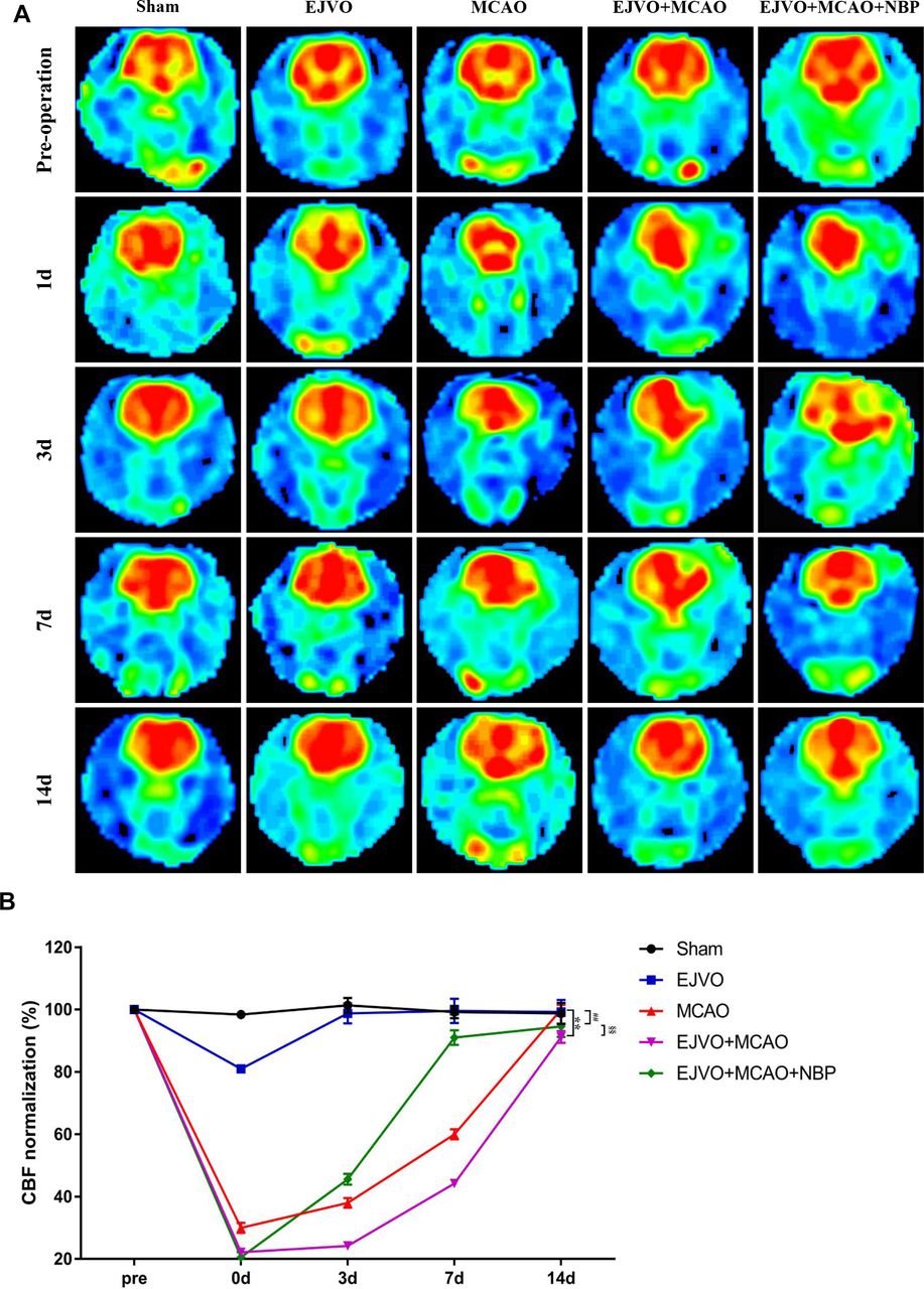
Cerebral venous circulation disturbance delayed recovery of CBF after MCAO, and NBP promoted CBF restoration. (A) CBF maps were generated from ASL and EJVO +MCAO group showed a lower CBF than MCAO group. NBP treatment could accelerate CBF recovery after rats underwent MCAO operation plus with EJVO. (B) Quantitative analysis showing changes of CBF in infarct area from first to 14th day. data are presented as mean±SD; n=5 per group. **P<0.01, EJVO +MCAO vs Sham; ##p<0.01, MCAO vs sham; §§p<0.01, EJVO +MCAO + NBP vs EJVO +MCAO. CBF, cerebral blood flow; EJVO, external jugular vein occlusion; MACO, middle cerebral artery occlusion; NBP, Dl-3-n-butylphthalide.
- Figure 7
Cerebral venous circulation disturbance aggravate the damage of blood brain barrier, and NBP could reduce BBB damage in EJVO +MCAO+ NBP group. (A) Representative images showing EB extravasation from first to 14th day after operation in the sham, EJVO, MCAO, EJVO +MCAO and EJVO +MCAO+ NBP groups. (B) Quantification analysis of absorbance of EB extravasation. (C) Quantification analysis of ipsilateral brain tissue water content. data are presented as mean±SD; n=5 per group. ¤P<0.05, EJVO vs sham; *p<0.05, MCAO vs sham; **p<0.01, MCAO vs Sham; ##p<0.01, EJVO +MCAO vs sham; §§p<0.01, EJVO +MCAO + NBP vs sham; ※p<0.05, MCAO vs EJVO; &p<0.05, EJVO +MCAO vs EJVO; &&p<0.01, EJVO +MCAO vs EJVO; ∫∫p<0.05, EJVO +MCAO vs MCAO; $p<0.05, EJVO +MCAO + NBP vs EJVO +MCAO; $$p<0.01, EJVO +MCAO + NBP vs EJVO +MCAO. Scale bar=5 mm. BBB, blood-brain barrier; EB, Evans blue; EJVO, external jugular vein occlusion; MACO, middle cerebral artery occlusion; NBP, Dl-3-n-butylphthalide.
- Figure 8
Cerebral venous circulation disturbance aggravated pericytes loss, and NBP treatment could protect pericyte from severe loss in rats of MCAO accompanied by EJVO. (A) the expression of pericytes labelled by colocalisaton of PDGFRβ (green) and desmin (red) in the ischaemic area or corresponding region. (B, C) Expression of PDGFRβ and desmin detected by Western blot. (D) Quantification analysis of PDGFRβ analysed by immunofluorescence staining. (E) quantification analysis of desmin analysed by immunofluorescence staining. (F, H) Quantification analysis of PDGFRβ analysed by Western blot. (G, I) Quantification analysis of desmin analysed by Western blot. Data are presented as mean±SD, n=5 per group for immunofluorescence staining and n=3 per group for Western bolt. *P<0.05, MCAO vs sham; **p<0.01, MCAO vs sham; #p<0.05, EJVO +MCAO vs Sham; ##p<0.01, EJVO +MCAO vs sham; §p<0.05, EJVO +MCAO + NBP vs sham; §§p<0.01, EJVO +MCAO + NBP vs sham; ※p<0.05, MCAO vs EJVO; ※※p<0.01, MCAO vs EJVO; &p<0.05, EJVO +MCAO vs EJVO; &&p<0.01, EJVO +MCAO vs EJVO; ∫∫p<0.05, EJVO +MCAO vs MCAO; $p<0.05, EJVO +MCAO + NBP vs EJVO +MCAO; $$p<0.01, EJVO +MCAO + NBP vs EJVO +MCAO. Scale bar=50 µm. EJVO, external jugular vein occlusion; MACO, middle cerebral artery occlusion; NBP, Dl-3-n-butylphthalide.
- Figure 9
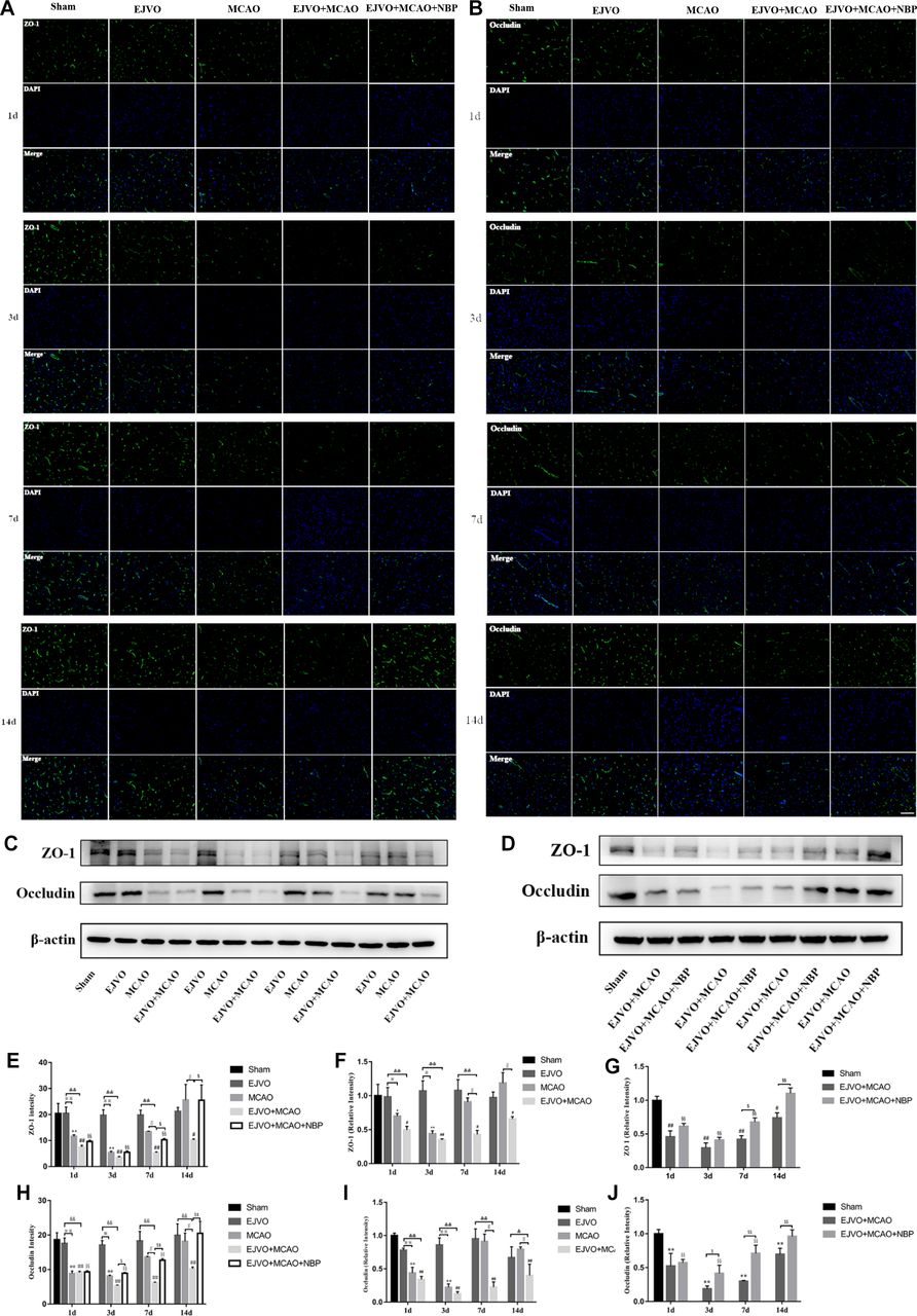
Cerebral venous circulation disturbance accelerate BBB damage, and NBP attenuated damage of BBB caused by MCAO plus with EJVO. (A) Expression of ZO-1 in the ischaemic area or corresponding region measured by immunofluorescence staining. (B) Expression of occludin in the ischaemic area or corresponding region measured by immunofluorescence staining. (C, D) Expression of ZO-1 and occludin detected by Western blot in the ischaemic brain tissue or corresponding area in the sham or EJVO group. (E) Quantification analysis of ZO-1 analysed by immunofluorescence staining. (F, G) Quantification analysis of ZO-1 analysed by Western blot. (H) Quantification analysis of occludin analysed by immunofluorescence staining. (I, J) Quantification analysis of occludin analysed by Western blot. data are presented as mean±SD, n=5 per group for immunofluorescence staining and n=3 per group for Western bolt. *P<0.05, MCAO vs sham; **p<0.01, MCAO vs sham; #p<0.05, EJVO +MCAO vs Sham; ##p<0.01, EJVO +MCAO vs sham; §§P<0.01, EJVO +MCAO + NBP vs sham; ※p<0.05, MCAO vs EJVO; ※※p<0.01, MCAO vs EJVO; &p<0.05, EJVO +MCAO vs EJVO; &&p<0.01, EJVO +MCAO vs EJVO; ∫∫p<0.05, EJVO +MCAO vs MCAO; $p<0.05, EJVO +MCAO + NBP vs EJVO +MCAO; $$p<0.01, EJVO +MCAO + NBP vs EJVO +MCAO. Scale bar=50 µm. BBB, blood-brain barrier; EJVO, external jugular vein occlusion; MACO, middle cerebral artery occlusion; NBP, Dl-3-n-butylphthalide.


