
Main menu

  • Online first
    • Online first
  • Current issue
    • Current issue
  • Archive
    • Archive
  • Submit a paper
    • Online submission site
    • Instructions for authors
  • About the journal
    • About the journal
    • Editorial board
    • Instructions for authors
    • FAQs
    • Chinese Stroke Association
  • Help
    • Contact us
    • Feedback form
    • Reprints
    • Permissions
    • Advertising
  • BMJ Journals

User menu

  • Login

Search

  • Advanced search
  • BMJ Journals
  • Login
  • Facebook
  • Twitter
Stroke and Vascular Neurology

Advanced Search

  • Online first
    • Online first
  • Current issue
    • Current issue
  • Archive
    • Archive
  • Submit a paper
    • Online submission site
    • Instructions for authors
  • About the journal
    • About the journal
    • Editorial board
    • Instructions for authors
    • FAQs
    • Chinese Stroke Association
  • Help
    • Contact us
    • Feedback form
    • Reprints
    • Permissions
    • Advertising
Open Access

Lesion-symptom mapping with NIHSS sub-scores in ischemic stroke patients

Deepthi Rajashekar, Matthias Wilms, M Ethan MacDonald, Serena Schimert, Michael D Hill, Andrew Demchuk, Mayank Goyal, Sean P Dukelow, Nils Daniel Forkert
DOI: 10.1136/svn-2021-001091 Published 27 April 2022
Deepthi Rajashekar
1 Biomedical Engineering Graduate Program, Hotchkiss Brain Institute, University of Calgary, Calgary, Alberta, Canada
2 Department of Radiology, Cumming School of Medicine, University of Calgary, Calgary, Alberta, Canada
3 Hotchkiss Brain Institute, University of Calgary, Calgary, Alberta, Canada
  • Find this author on Google Scholar
  • Find this author on PubMed
  • Search for this author on this site
  • ORCID record for Deepthi Rajashekar
Matthias Wilms
2 Department of Radiology, Cumming School of Medicine, University of Calgary, Calgary, Alberta, Canada
  • Find this author on Google Scholar
  • Find this author on PubMed
  • Search for this author on this site
M Ethan MacDonald
2 Department of Radiology, Cumming School of Medicine, University of Calgary, Calgary, Alberta, Canada
4 Department of Clinical Neurosciences, University of Calgary, Calgary, Alberta, Canada
5 Department of Electrical and Computer Engineering, University of Calgary, Calgary, Alberta, Canada
  • Find this author on Google Scholar
  • Find this author on PubMed
  • Search for this author on this site
Serena Schimert
2 Department of Radiology, Cumming School of Medicine, University of Calgary, Calgary, Alberta, Canada
4 Department of Clinical Neurosciences, University of Calgary, Calgary, Alberta, Canada
  • Find this author on Google Scholar
  • Find this author on PubMed
  • Search for this author on this site
Michael D Hill
2 Department of Radiology, Cumming School of Medicine, University of Calgary, Calgary, Alberta, Canada
3 Hotchkiss Brain Institute, University of Calgary, Calgary, Alberta, Canada
4 Department of Clinical Neurosciences, University of Calgary, Calgary, Alberta, Canada
6 Department of Community Health Sciences, Cumming School of Medicine, University of Calgary, Calgary, Alberta, Canada
7 Department of Medicine, Cumming School of Medicine, University of Calgary, Calgary, Alberta, Canada
  • Find this author on Google Scholar
  • Find this author on PubMed
  • Search for this author on this site
Andrew Demchuk
2 Department of Radiology, Cumming School of Medicine, University of Calgary, Calgary, Alberta, Canada
7 Department of Medicine, Cumming School of Medicine, University of Calgary, Calgary, Alberta, Canada
  • Find this author on Google Scholar
  • Find this author on PubMed
  • Search for this author on this site
Mayank Goyal
2 Department of Radiology, Cumming School of Medicine, University of Calgary, Calgary, Alberta, Canada
7 Department of Medicine, Cumming School of Medicine, University of Calgary, Calgary, Alberta, Canada
  • Find this author on Google Scholar
  • Find this author on PubMed
  • Search for this author on this site
Sean P Dukelow
3 Hotchkiss Brain Institute, University of Calgary, Calgary, Alberta, Canada
4 Department of Clinical Neurosciences, University of Calgary, Calgary, Alberta, Canada
  • Find this author on Google Scholar
  • Find this author on PubMed
  • Search for this author on this site
Nils Daniel Forkert
2 Department of Radiology, Cumming School of Medicine, University of Calgary, Calgary, Alberta, Canada
3 Hotchkiss Brain Institute, University of Calgary, Calgary, Alberta, Canada
4 Department of Clinical Neurosciences, University of Calgary, Calgary, Alberta, Canada
8 Alberta Children's Hospital Research Institute, University of Calgary, Calgary, AB, Canada
  • Find this author on Google Scholar
  • Find this author on PubMed
  • Search for this author on this site
  • ORCID record for Nils Daniel Forkert
  • Article
  • Figures & Data
  • eLetters
  • Info & Metrics
  • PDF
Loading

Article Figures & Data

Figures

  • Tables
  • Supplementary Materials
  • Additional Files
  • Figure 1
    • Download figure
    • Open in new tab
    • Download powerpoint
    Figure 1

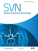
    Overlap of all 180 subject lesions on the MIPLAB atlas in radiological convention. The maximum occurrence of lesions in this data sample (shown in red) is in the MCA territory. Implying, LSM (in this work) focuses on determining structure–function relationships of brain regions with respect to the NIHSS subdomains. LSM, lesion-symptom mapping; MCA, middle cerebral artery; NIHSS, National Institutes of Health Stroke Scale.

  • Figure 2
    • Download figure
    • Open in new tab
    • Download powerpoint
    Figure 2

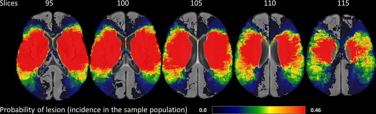
    LSM analyses for the total NIHSS score (total map): higher eloquence scores indicate regions critically associated with the 48-hour NIHSS. Results show that even a small lesion suffices to result in severe deficit in the left hemispheric regions. LSM, lesion-symptom mapping; NIHSS, National Institutes of Health Stroke Scale.

  • Figure 3
    • Download figure
    • Open in new tab
    • Download powerpoint
    Figure 3

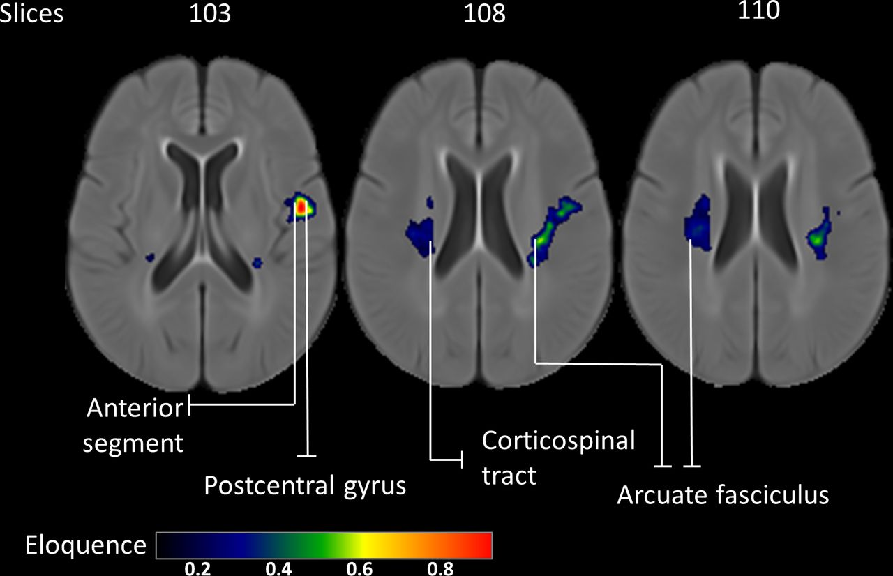
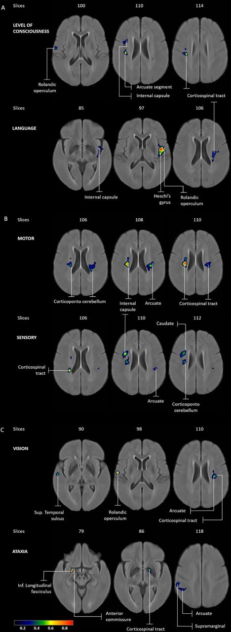
    LSM analyses for score-based NIHSS grouping: (A) consciousness and language—all regions except the left insula and left anterior segment are critical in both consciousness (top) and language LSM (below); (B) motor and sensory—bilateral critical association of the arcuate and corticospinal tract in motor map (top), whereas in the sensory LSM (below) these tracts are critical only in the right hemisphere; (C) vision and neglect—with the exception of arcuate, the vision and sensory LSMs have distinct critical regions. LSM, lesion-symptom mapping; NIHSS, National Institutes of Health Stroke Scale.

  • Figure 4
    • Download figure
    • Open in new tab
    • Download powerpoint
    Figure 4

    LSM analyses for anatomy-based NIHSS grouping: (A) consciousness LSM (top) shows predominantly right hemispheric regions while language LSM (below) shows left hemispheric brain regions; (B) bilateral critical association of the arcuate and corticospinal tract is identified in the motor LSM (top) whereas the sensory LSM (below) shows critical regions only in the right hemisphere; (C) vision LSM (top) shows bilateral critical regions while the ataxia LSM shows right hemispheric dominance. LSM, lesion-symptom mapping; NIHSS, National Institutes of Health Stroke Scale.

  • Figure 5
    • Download figure
    • Open in new tab
    • Download powerpoint
    Figure 5

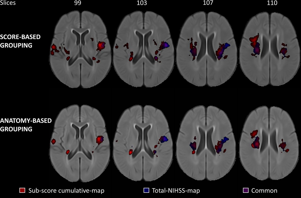
    Comparison of the cumulative LSM (red) and the total LSM (blue) overlaid on the MIPLAB atlas. The cumulative map does not only contain most of the critical regions seen in the total LSM but reveals more structure–function relationships by utilising NIHSS sub-score information. Brain regions that are common to both, the total NIHSS total map and the sub-score cumulative map, are shown in violet. LSM, lesion-symptom mapping; NIHSS, National Institutes of Health Stroke Scale.

Tables

  • Figures
  • Supplementary Materials
  • Additional Files
  • Table 1

    Summary of the two schemes used to categorise NIHSS sub-scores assessed at 48 hours (N=180, 84 men).

    CategoryScore-based groupingAnatomy-based grouping
    Level of consciousness1a+1 b+1 c1a+2
    Language9+101b+1 c+9 + 10
    Motor4+5 a+5 b+6 a+6 b+74+5 a+5 b+6 a+6b
    Sensory88+11
    Vision2+33
    Ataxia–7
    Neglect11–
    • The numbers correspond to the standard definition of the 11-item modified NIHSS scale.10 11 Anatomy-based grouping considers neuroanatomy to group the NIHSS components, while score-based grouping follows the design of the NIHSS scale alone.

    • NIHSS, National Institutes of Health Stroke Scale.

Supplementary Materials

  • Figures
  • Tables
  • Additional Files
  • Supplementary data

    [svn-2021-001091supp001.pdf]

Additional Files

  • Figures
  • Tables
  • Supplementary Materials
  • Supplementary Data

    This web only file has been produced by the BMJ Publishing Group from an electronic file supplied by the author(s) and has not been edited for content.

    • Data supplement 1
PreviousNext
Back to top
Vol 7 Issue 2 Table of Contents
Stroke and Vascular Neurology: 7 (2)
  • Table of Contents
  • Table of Contents (PDF)
  • About the Cover
  • Index by author
  • Front Matter (PDF)
Email

Thank you for your interest in spreading the word on Stroke and Vascular Neurology.

NOTE: We only request your email address so that the person you are recommending the page to knows that you wanted them to see it, and that it is not junk mail. We do not capture any email address.

Enter multiple addresses on separate lines or separate them with commas.
Lesion-symptom mapping with NIHSS sub-scores in ischemic stroke patients
(Your Name) has sent you a message from Stroke and Vascular Neurology
(Your Name) thought you would like to see the Stroke and Vascular Neurology web site.
CAPTCHA
This question is for testing whether or not you are a human visitor and to prevent automated spam submissions.
Print
Alerts
Sign In to Email Alerts with your Email Address
Citation Tools
Lesion-symptom mapping with NIHSS sub-scores in ischemic stroke patients
Deepthi Rajashekar, Matthias Wilms, M Ethan MacDonald, Serena Schimert, Michael D Hill, Andrew Demchuk, Mayank Goyal, Sean P Dukelow, Nils Daniel Forkert
Stroke and Vascular Neurology Apr 2022, 7 (2) 124-131; DOI: 10.1136/svn-2021-001091

Citation Manager Formats

  • BibTeX
  • Bookends
  • EasyBib
  • EndNote (tagged)
  • EndNote 8 (xml)
  • Medlars
  • Mendeley
  • Papers
  • RefWorks Tagged
  • Ref Manager
  • RIS
  • Zotero
Cite This
  • APA
  • Chicago
  • Endnote
  • MLA
Loading
Lesion-symptom mapping with NIHSS sub-scores in ischemic stroke patients
Deepthi Rajashekar, Matthias Wilms, M Ethan MacDonald, Serena Schimert, Michael D Hill, Andrew Demchuk, Mayank Goyal, Sean P Dukelow, Nils Daniel Forkert
Stroke and Vascular Neurology Apr 2022, 7 (2) 124-131; DOI: 10.1136/svn-2021-001091
Download PDF

Share
Lesion-symptom mapping with NIHSS sub-scores in ischemic stroke patients
Deepthi Rajashekar, Matthias Wilms, M Ethan MacDonald, Serena Schimert, Michael D Hill, Andrew Demchuk, Mayank Goyal, Sean P Dukelow, Nils Daniel Forkert
Stroke and Vascular Neurology Apr 2022, 7 (2) 124-131; DOI: 10.1136/svn-2021-001091
Reddit logo Twitter logo Facebook logo Mendeley logo
Respond to this article
  • Tweet Widget
  • Facebook Like
  • Google Plus One
  • Article
    • Abstract
    • Introduction
    • Materials and methods
    • Methods
    • Results
    • Discussion
    • Conclusion
    • Data availability statement
    • Ethics statements
    • Acknowledgments
    • Footnotes
    • References
  • Figures & Data
  • eLetters
  • Info & Metrics
  • PDF

Related Articles

Cited By...

More in this TOC Section

  • Learning curve and embolisation strategy in single-stage surgery combined embolisation and microsurgery for brain arteriovenous malformations: results from a nationwide multicentre prospective registry study
  • Stepwise improvement in intracerebral haematoma expansion prediction with advanced imaging: a comprehensive comparison of existing scores
  • Thrombus iodine-based perviousness is associated with recanalisation and functional outcomes in endovascular thrombectomy
Show more Original research

Similar Articles


CONTENT

  • Latest content
  • Current issue
  • Archive
  • eLetters
  • Sign up for email alerts
  • RSS

JOURNAL

  • About the journal
  • Editorial board
  • Recommend to librarian
  • Chinese Stroke Association

AUTHORS

  • Instructions for authors
  • Submit a paper
  • Track your article
  • Open Access at BMJ

HELP

  • Contact us
  • Reprints
  • Permissions
  • Advertising
  • Feedback form

© 2025 Chinese Stroke Association