

Article Figures & Data
Figures
- Figure 1
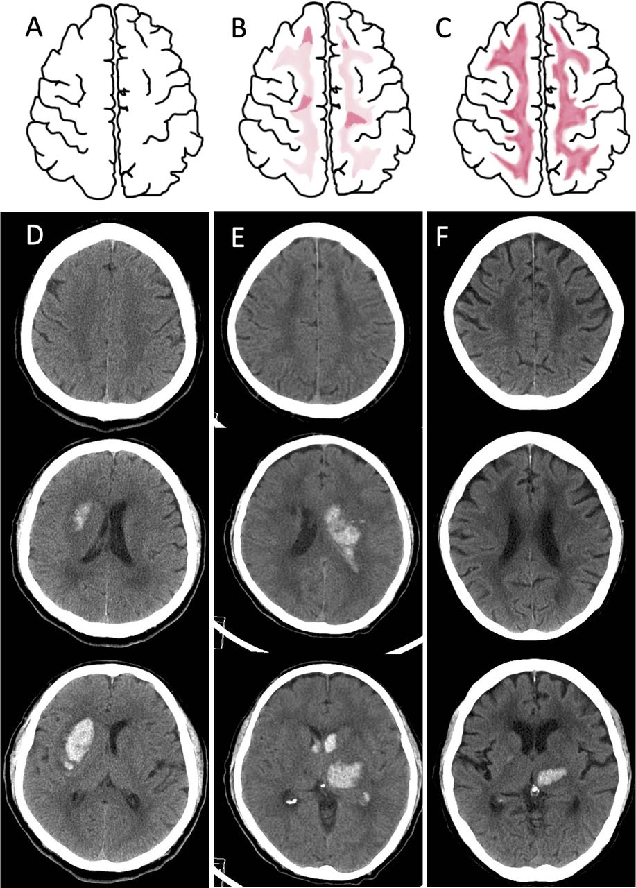
Schematic and neuroimaging demonstration of prominent juxtacortical WML. (A–C) Schematic diagrams of the WML at the level of CS. (A) A normal CS without WML. (B) Pattern 1: bilateral vague hypodensity change in CS with patchy areas of low densities inside, involving the juxtacortical region. (C) Pattern 2: bilateral diffuse hypodensity change in CS, extending to the juxtacortical region, delineating a sharp interface of grey–white matter junction. (D–F) CT scans of 3 different patients of ICH. (D) A case of right putaminal haemorrhage with negative NOTCH3 p.R544C mutation was scored 1 and 2 for frontal and parietal lobe WML but without WML in the CS. (E) A case of left thalamic haemorrhage with positive NOTCH3 p.R544C mutation, was scored 2 for frontal and parietal lobe WML, with pattern 1 change in CS. (F) A case of left thalamic haemorrhage with positive NOTCH3 p.R544C mutation was scored 2 for frontal and parietal lobe WML, with pattern 2 change in the CS. Note that the ventricular-level white matter also reveals extensive WML reaching the grey–white matter interface. CS, centrum semiovale; ICH, intracerebral haemorrhage; WML, white matter lesion.
- Figure 2
Flowchart of participant recruitment. FSGC, Formosan Stroke Genetic Consortium; ICH, intracerebral haemorrhage; NTUH, National Taiwan University Hospital; SAH, subarachnoid haemorrhage; TIA, transient ischaemic attack.
- Figure 3
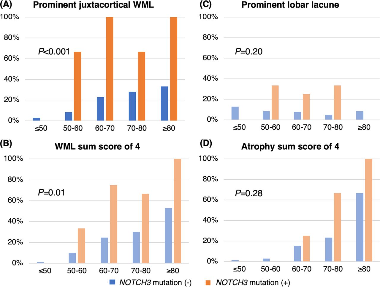
The presence of CT features across different age groups between patients with and without NOTCH3 p.R544C mutation. (A) Prominent juxtacortical white matter lesion (WML), (B) maximum WML score, (C) prominent lobar lacune and (D) maximum cerebral atrophy score. P value was derived from the Cochrane-Mantel-Haenszel test.


Tables
- Table 1
Clinical characteristics between patients with ICH with and without NOTCH3 p.R544C mutation
NOTCH3 mutation (−) n=286 NOTCH3 mutation (+) n=13 P value Age 61.3±14.7 64.2±12.1 0.44 Male sex 194 (67.8%) 8 (61.5%) 0.64 Medical history Hypertension 263 (92.0%) 11 (84.6%) 0.30 Diabetes mellitus 88 (31.0%) 5 (38.5%) 0.55 Hyperlipidaemia 89 (31.1%) 9 (69.2%) 0.004 Smoking 104 (36.6%) 3 (23.1%) 0.39 Heart disease 68 (23.9%) 1 (7.7%) 0.31 Previous ischaemic stroke 29 (10.1%) 2 (15.4%) 0.65 Previous ICH 27 (9.4%) 3 (23.1%) 0.16 Parental stroke 72 (25.4%) 4 (30.8%) 0.75 Sibling stroke 37 (13.1%) 4 (30.8%) 0.09 ICH location 0.39 Thalamus 77 (26.9%) 6 (46.2%) Basal ganglia 106 (37.1%) 3 (23.1%) Cortical-subcortical 55 (19.2%) 1 (7.7%) Infratentorium 46 (16.1%) 3 (23.1%) Multiple or others 2 (1.0%) 0 (0%) ICH size, mL 14.2±17.9 13.4±17.6 0.30 IVH 90 (31.7%) 4 (30.8%) 0.99 Admission status Glasgow coma scale 12.6±3.5 13.0±3.2 0.58 Systolic blood pressure 182.7±37.8 185.0±29.9 0.76 Diastolic blood pressure 101.7±25.0 100.5±14.2 0.96 Heart rate 86.3±17.8 86.0±23.2 0.36 NIHSS 13.0±9.5 13.1±11.5 0.77 ICH score 1.2±1.2 1.2±1.1 0.96 Discharge status Length of stay, days 15.5±14.4 10.9±5.7 0.48 NIHSS at discharge 10.1±11.6 13.8±14.0 0.37 Change of NIHSS −3.3±8.3 0.7±7.1 0.02 Barthel index 59.6±36.8 39.2±44.9 0.13 Modified Rankin scale 2.9±1.6 3.5±1.9 0.20 Numbers in bold indicating statistical significance (P<0.05).
ICH, intracerebral haemorrhage; IVH, intraventricular haemorrhage; NIHSS, National Institute of Health Stroke Scale.
- Table 2
CT features distinguishing patients with ICH with and without NOTCH3 p.R544C mutation
NOTCH3 mutation (−), n=286 NOTCH3 mutation (+), n=13 P value Unadjusted OR (95% CI) Demographic-adjusted OR (95% CI)* Anterior WML score 0.9±0.8 1.4±0.9 0.03 2.23 (1.05 to 4.74) 2.84 (1.22 to 6.60) Posterior WML score 0.7±0.8 1.4±0.9 0.01 2.44 (1.24 to 4.81) 3.56 (1.60 to 7.91) Total WML score 1.6±1.5 2.8±1.7 0.02 1.61 (1.11 to 2.35) 1.99 (1.27 to 3.12) Prominent juxtacortical WML 47 (16.4%) 9 (69.2%) <0.001 10.6 (3.31 to 34.3) 20.9 (4.94 to 88.6) Pattern 1 26 (55.3%) 4 (44.4%) 0.72 Pattern 2 21 (44.7%) 5 (55.6%) Anterior temporal lobe WML 1 (0.35%) 0 (0%) 0.83 NA NA External capsule WML 33 (11.5%) 6 (46.2%) <0.001 6.56 (2.14 to 20.1) 8.28 (2.27 to 30.2) Cortical atrophy score 0.7±0.7 1.0±0.8 0.24 1.56 (0.77 to 3.17) 1.85 (0.69 to 4.96) Central atrophy score 0.9±0.8 1.2±1.0 0.25 1.51 (0.79 to 2.91) 1.75 (0.74 to 4.14) Total atrophy score 1.6±1.5 2.2±1.7 0.23 1.28 (0.89 to 1.83) 1.44 (0.87 to 2.41) Presence of lacune 125 (43.7%) 8 (61.5%) 0.26 1.99 (0.66 to 5.98) 2.15 (0.72 to 6.42) Number of lacune 1.2±1.9 1.9±2.1 0.13 1.18 (0.95 to 1.47) 1.21 (0.96 to 1.51) Prominent lobar lacune 25 (8.7%) 3 (23.1%) 0.11 3.42 (0.94 to 12.5) 3.91 (1.03 to 14.8) Numbers in bold indicating statistical significance (P<0.05).
*Adjusted by age, sex and hyperlipidaemia.
WML, white matter lesion.
- Table 3
Performance of clinical and CT features in predicting NOTCH3 p.R544C mutation
aOR (95% CI)* AUC-ROC (95% CI) P value † Total WML score 0.79 (0.37 to 1.66) 0.69 (0.52 to 0.86) 0.047 External capsule WML 1.70 (0.39 to 7.33) 0.67 (0.53 to 0.82) 0.141 Prominent lobar lacune 2.32 (0.53 to 10.13) 0.57 (0.45 to 0.69) 0.038 Prominent juxtacortical WML 20.4 (1.54 to 271.0) 0.76 (0.63 to 0.90) – Numbers in bold indicating statistical significance (P<0.05).
*Simultaneously adjusted with these three imaging markers.
†Compared with prominent juxtacortical WML alone.
aOR, adjusted OR; AUC-ROC, area under the receive operating characteristic curve; WML, white matter lesion.
- Table 4
Comparison between patients with NOTCH3 p.R544C mutation with and without prominent juxtacortical white matter lesion
NOTCH3 mutation, juxtacortical WML (+) NOTCH3 mutation, juxtacortical WML (−) Non-mutation, juxtacortical WML (+) Number 9 4 47 Age 67.3±10.1 57.3±14.6 71.8±11.7 Male sex 6 (66.7%) 2 (50.0%) 32 (68.1%) ICH location Thalamus 4 (44.4%) 2 (50.0%) 19 (40.4%) Basal ganglia 2 (22.2%) 1 (25.0%) 8 (17.0%) Cortical-subcortical 1 (11.1%) 0 (0.0%) 11 (23.4%) Infratentorium 2 (22.2%) 1 (25.0%) 9 (19.1%) ICH size, mL 13.4±17.7 13.4±21.3 13.0±17.5 IVH 3 (33.3%) 1 (25.0%) 21 (44.7%) CT features Anterior WML score 1.9±0.3 0.3±0.5 1.9±0.3 Posterior WML score 1.9±0.3 0.3±0.5 1.9±0.3 Total WML score 3.8±0.4 0.5±1.0 3.8±0.5 Anterior temporal lobe WML 0 (0%) 0 (0%) 1 (2.1%) External capsule WML 6 (66.7%) 0 (0%) 21 (44.7%) Cortical atrophy score 1.2±0.8 0.5±0.6 1.2±0.7 Central atrophy score 1.4±0.9 0.5±1.0 1.6±0.6 Total atrophy score 2.7±1.7 1.0±1.4 2.9±1.1 Presence of lacune 7 (77.8%) 1 (25.0%) 30 (63.8%) Number of lacune 2.7±2.2 0.3±0.5 2.3±2.8 Prominent lobar lacune 2 (22.2%) 1 (25.0%) 8 (17.0%) ICH, intracerebral haemorrhage; IVH, intraventricular haemorrhage; WML, white matter lesion.
- Table 5
Comparison of the neuroimaging features between patients who had received both the CT and MRI scans
N=145 CT MRI Kappa values (95% CI) Anterior WML≥2 46 (31.7%) 60 (41.4%) 0.74 (0.62 to 0.85) Posterior WML ≥2 43 (29.7%) 67 (46.2%) 0.63 (0.51 to 0.75) Prominent juxtacortical WML 33 (22.9%) 44 (30.6%) 0.81 (0.70 to 0.91) Anterior temporal lobe WML 0 (0%) 1 (0.7%) 0.66 (0.04 to 1.00) External capsule WML 23 (16.0%) 22 (15.3%) 0.97 (0.92 to 1.00) Presence of lacune 70 (48.6%) 76 (52.8%) 0.81 (0.71 to 0.90) WML, white matter lesion.


