

Article Figures & Data
- Figure 1
Sankey diagrams showing the distribution and changes in mRS score at enrolment and at censoring time. (A) The figure consisted of two bars and one flow in between. These two bars show the distribution of mRS scores at enrolment and at censoring time in orange. The width and value of each flow were calculated according to the sample size showing the dominant or subordinate contributions to the overall flow that were similar in other panels. (B–D) The mRS scores at enrolment and at censoring time were stratified by the number of prospective haemorrhages (B), crossing the axial midpoint or not (C), and presence of DVA or not (D). The Sankey diagrams are illustrated using Tableau Desktop (V.8.3). DVA, developmental venous anomaly; mRS, modified Rankin Scale.
- Figure 2
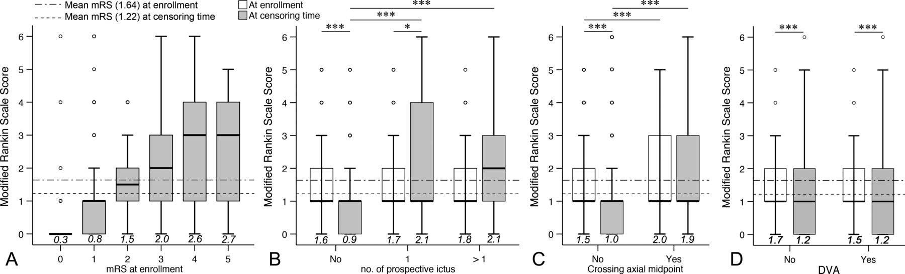
Boxplots showing the distribution of mRS scores at enrolment and at censoring time based on risk factors. (A) The boxplot shows the mRS score at censoring time stratified by mRS score at enrolment. (B–D) The outcome was significantly better in patients without prospective ictus than in patients with one (Z=−7.398, p<0.001) or more (Z=−5.414, p<0.001) ictus (B). Patients without lesions crossing the axial midpoint had significantly better outcomes than patients with lesions crossing the axial midpoint (Z=−6.352, p<0.001) (C). However, there was no significance between the outcomes in patients with or without DVA (Z=−0.384, p=0.701) (D). *P<0.05, ***P<0.001. Circles indicate the outliers; values in italic above the horizontal axis indicate the mean values of each boxplot. DVA, developmental venous anomaly; mRS, modified Rankin Scale.


- Table 1
Baseline and clinical data of brainstem cavernous malformations stratified by neurological outcome
Variable (%) Overall, N=698 Improved (mRS=0), n=334 Unchanged, n=293 Worsened, n=71 P value Female 321 (46.0) 154 (46.1) 132 (45.1) 35 (49.3) 0.811† Mean age at enrolment, years 38.3±13.8 36.8±13.3 39.5±13.8 40.6±15.2 0.017*‡ Hypertension 96 (13.8) 50 (15.0) 38 (13.0) 8 (11.3) 0.625† Mean duration of symptoms, months 21.3±44.7 16.4±37.9 24.5±46.3 30.8±62.5 0.013*‡ Patients with prior haemorrhage 680 (97.4) 322 (96.4) 288 (98.3) 70 (98.6) 0.267† Mean number of prior haemorrhage 1.4±0.8 1.3±0.8 1.5±0.9 1.6±0.8 0.003*‡ Haemorrhage at enrolment 508 (72.8) 228 (68.3) 223 (76.1) 57 (80.3) 0.029*† FND at enrolment 589 (84.4) 265 (79.3) 262 (89.4) 62 (87.3) 0.002*† Midbrain location 154 (22.1) 69 (20.7) 65 (22.2) 20 (28.2) 0.699† Pontine location 442 (63.3) 216 (64.7) 186 (63.5) 40 (56.3) Medullary location 102 (14.6) 49 (14.7) 42 (14.3) 11 (15.5) Crossing axial midpoint 174 (24.9) 70 (21.0) 71 (24.2) 33 (46.5) <0.001*† Developmental venous anomaly 237 (34.0) 106 (31.7) 98 (33.4) 33 (46.5) 0.057† Perilesional oedema 342 (49.0) 172 (51.5) 132 (45.1) 38 (53.5) 0.198† Zabramski type I 190 (27.2) 97 (29.0) 70 (23.9) 23 (32.4) 0.010*† Zabramski type II 326 (46.7) 135 (40.4) 156 (53.2) 35 (49.3) Zabramski type III/IV 182 (26.1) 102 (30.5) 67 (22.9) 13 (18.3) Mean lesion size, cm¶ 1.5±0.7 1.4±0.6 1.6±0.6 1.9±0.7 <0.001*‡ Size >1.5 cm 299 (42.8) 116 (34.7) 135 (46.1) 48 (67.6) <0.001*† Superficial-seated 395 (56.6) 173 (51.8) 171 (58.4) 51 (71.8) 0.003*† Moderate-seated 208 (29.8) 102 (30.5) 93 (31.7) 13 (18.3) Deep-seated 95 (13.6) 59 (17.7) 29 (9.9) 7 (9.9) mRS score at enrolment 0.363§ 0 47 (6.7) 41 (12.3) 0 6 (8.5) 1 400 (57.3) 153 (45.8) 210 (71.7) 37 (52.1) 2 104 (14.9) 52 (15.6) 38 (13.0) 14 (19.7) 3 66 (9.5) 39 (11.7) 17 (5.8) 10 (14.1) 4 68 (9.7) 38 (11.4) 26 (8.9) 4 (5.6) 5 13 (1.9) 11 (3.3) 2 (0.7) 0 mRS score at censored <0.001*§ 0 233 (33.4) 233 (69.8) 0 0 1 283 (40.5) 70 (21.0) 210 (71.7) 3 (4.2) 2 71 (10.2) 17 (5.1) 38 (13.0) 16 (22.5) 3 44 (6.3) 10 (3.0) 17 (5.8) 17 (23.9) 4 49 (7.0) 4 (1.2) 26 (8.9) 19 (26.8) 5 6 (0.9) 0 2 (0.7) 4 (5.6) 6 12 (1.7) 0 0 12 (16.9) Mean follow-up duration, months 56.9±35.3 69.0±28.5 49.8±37.6 29.4±31.3 <0.001*‡ Haemorrhage-free survival time, months 48.7±36.0 61.4±32.2 41.9±36.6 17.0±21.0 <0.001*‡ Patients with prospective bleeding 167 (23.9) 48 (14.4) 56 (19.1) 63 (88.7) <0.001*† Mean number of prospective bleeding 0.3±0.7 0.2±0.5 0.3±0.6 1.2±0.8 <0.001*‡ Patients under observation 514 (73.6) 301 (90.1) 188 (64.2) 25 (35.2) <0.001*† Patients receiving radiosurgery 14 (2.0) 5 (1.5) 7 (2.4) 2 (2.8) Patients receiving surgery 170 (24.4) 28 (8.4) 98 (33.4) 44 (62.0) Means are given with SD.
Bold indicates statistical significance.
*P<0.05.
†χ2 test.
‡One-way analysis of variance.
§Kruskal-Wallis test.
¶Lesion size was expressed as the lesion equivalent diameter (abc)1/3, where a, b and c represent the maximal diameters (length, width and height) measured on axial, sagittal and coronal MRI scans.
FND, focal neurological deficit; mRS, modified Rankin Scale.
- Table 2
Risk factors for worsened neurological function in brainstem cavernous malformations
Variable (%) n Worsened (n=71, %) Univariate†
RR (95% CI)P value Multivariate†‡
RR (95% CI)P value Male 377 36 (9.5) Reference Reference Female 321 35 (10.9) 1.159 (0.709 to 1.894) 0.555 1.074 (0.634 to 1.819) 0.792 Age (per 1 year) 1.014 (0.995 to 1.032) 0.145 1.020 (1.000 to 1.041) 0.051 Without hypertension 602 63 (10.5) Reference Hypertension 96 8 (8.3) 0.778 (0.360 to 1.679) 0.522 Duration of symptoms (per 0.1 months) 1.004 (1.000 to 1.008) 0.064 1.004 (0.999 to 1.009) 0.101 Without prior haemorrhage 18 1 (5.6) Reference With prior haemorrhage 680 70 (10.3) 1.951 (0.256 to 14.882) 0.519 Without haemorrhage at enrolment 190 14 (7.4) Reference Haemorrhage at enrolment 508 57 (11.2) 1.589 (0.863 to 2.924) 0.137 Without FND at enrolment 109 9 (8.3) Reference FND at enrolment 589 62 (10.5) 1.307 (0.629 to 2.715) 0.473 mRS score at enrolment (per one score) 0.939 (0.756 to 1.168) 0.573 Lesion size (per 1 mm) 2.343 (1.643 to 3.341) <0.001* 1.263 (0.806 to 1.979) 0.309 Not crossing axial midpoint 524 38 (7.3) Reference Reference Crossing axial midpoint 174 33 (19.0) 2.993 (1.811 to 4.948) <0.001* 2.325 (1.332 to 4.060) 0.003* Without venous anomaly 461 38 (8.2) Reference Reference Developmental venous anomaly 237 33 (13.9) 1.801 (1.097 to 2.955) 0.020* 1.776 (1.037 to 3.041) 0.036* Depth (superficial-seated) 395 51 (12.9) 1.595 (1.074 to 2.367) 0.021* 1.194 (0.774 to 1.841) 0.423 Without oedema 356 33 (9.3) Reference Perilesional oedema 342 38 (11.1) 1.223 (0.748 to 2.001) 0.422 Zabramski classification 1.313 (0.935 to 1.846) 0.116 Type III/IV 182 13 (7.1) Reference Type II 326 35 (10.7) 1.564 (0.805 to 3.038) 0.187 Type I 190 23 (12.1) 1.790 (0.878 to 3.652) 1.790 Bold indicates statistical significance.
*P<0.05.
†Binary logistic regression (method: Enter); risk factors, entered into the multivariate binary logistic regression, included gender, age, duration of symptoms, lesion size, crossing axial midpoint, developmental venous anomaly and depth of the lesion.
‡Adjustment for duration of follow-up.
FND, focal neurological deficit; mRS, modified Rankin Scale; RR, relative risk.
- Table 3
Neurological outcome of untreated brainstem CMs
Study and year Study type Patients (n) Mean age, years Mean follow-up, years Patients with prospective ictus (%) Total number of ictus Annual haemorrhage rate, % Neurological outcome (%) Zimmerman et al 199130 Retrospective 8 NA 3–60 months 1 (12.5) 1 NA 7 (87.5) minor symptoms or asymptomatic; 1 (12.5) died of haemorrhage. Fritschi et al 199426 Retrospective 30 NA 3.0 NA NA NA 13 (43.3) normal; 7 mildly, 2 moderately and 2 severely disabled; 6 (20.0) died of CMs. Bouillot et al 199629 Retrospective 7 NA 5.6 NA NA 11.7 1 (14.3) improved; 4 (57.1) same; 2 (28.6) worsened. Porter et al 199920 Retrospective 12 NA NA NA NA NA 7 (58.3) improved/same; 5 (41.7) worsened; 1 (8.3) died of rebleeding. Kupersmith et al 200112 Retrospective 37 37.5 4.9 7 (18.9) 8 5.1 18 (48.6) improved; 14 (37.8) same; 3 (8.1) worsened; 2 (5.4) died of myocardial infarction. Esposito et al 200332 Retrospective 17 NA NA NA NA 2.5 11 (64.7) favourable; 5 (29.4) same; 1 (5.9) worsened. Tarnaris et al 200828 Retrospective 15 37.9 6.1 4 (26.7) 4 4.4 2 (13.3) improved; 5 (33.3) same; 8 (53.3) worsened; mean mRS score 2.07. Bhardwaj et al 200931 Retrospective 13‡ 12.7 4.4 1 (7.7) 1 ≈1.7 4 (30.8) improved; 8 (61.5) same (6 asymptomatic, 2 symptomatic); 1 (7.7) died. Chen et al 201121 Retrospective 20 38.1 3.7 7 (35.0) ≥11 NA Mean NIHSS score 1.7. Menon et al 201127 Retrospective 29† NA 4.0 17 (63.0) ≥41 ≥35.3 14 (51.9) improved/same; 13 (48.1) worsened; 1 (3.7) died of haemorrhage. Al-Holou et al 20128 Retrospective 15 les NA ≈3.5 5 les (33.3) 8 ≈15.2 per les NA Al-Shahi Salman et al 20122 Prospective 14 NA 5.0 NA NA NA 8 patients (57.1) had a second haemorrhage or focal neurological deficit. Li et al 201417 Prospective 85 12.7 4.7 37 (43.5) 47 11.7 22 (25.9) normal; 33 (38.8) improved; 32 (37.6) same; 20 (23.5) worsened. Li et al 201417 Prospective 331 36.2 6.5 131 (39.6) 185 13.6 95 (28.7) normal; 198 (59.8) improved; 109 (32.9) same; 24 (7.3) worsened; 3 (0.9) deaths. Gross et al 20165 Retrospective 18 lesion NA 3.3 NA 10 16.7 per lesion Permanent neurological morbidity of 45% for brainstem, thalamic and basal ganglia CMs. Horne et al 20163 Meta-analysis 575 NA NA 139 (24.2) 139 NA NA Zuurbier et al 201914 Prospective 34 NA NA NA 15 NA 15 events of haemorrhage or persistent or progressive focal neurological deficit. Present series Prospective 698 38.3 4.7 167 (23.9) 222 6.7 233 (33.4) normal; 334 (47.9) improved/mRS=0, 293 (42.0) same; 71 (10.2) worsened. *Two patients were lost to follow-up; 5 had one haemorrhage and 12 had more than two haemorrhages during follow-up.
†Seven patients had previous radiotherapy and one patient died of medulloblastoma.
CMs, cavernous malformations; les, lesions; mRS, modified Rankin Scale; NA, not available; NIHSS, National Institutes of Health Stroke Scale.
Supplementary Materials
Supplementary data
Additional Files
Supplementary Data
This web only file has been produced by the BMJ Publishing Group from an electronic file supplied by the author(s) and has not been edited for content.


