

Article Figures & Data
Figures
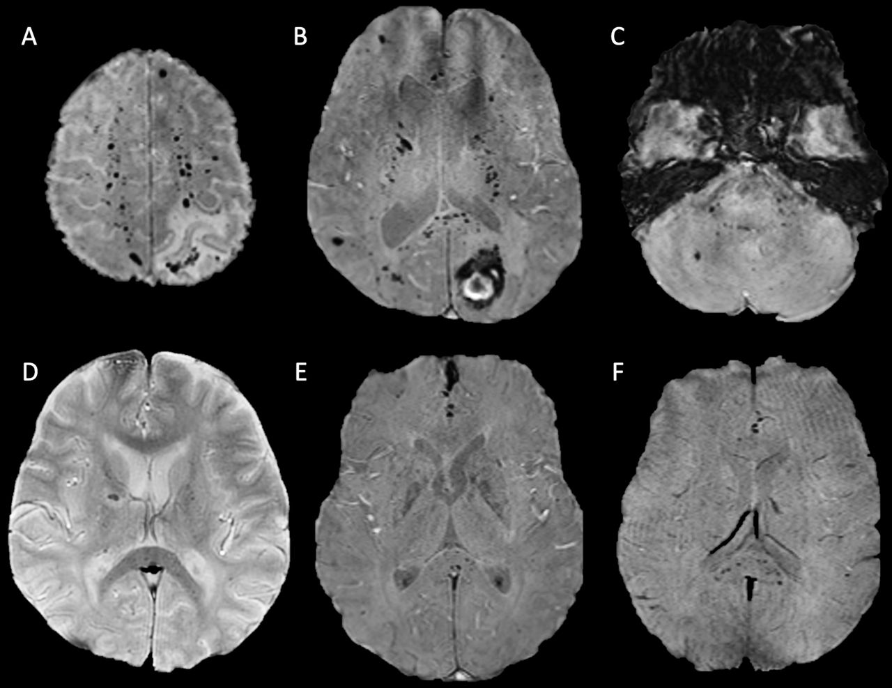
- Figure 1
Axial susceptibility-weighted MRI sequences of patient 4 (A–C) and patients 2 (D), 5 (E) and 10 (F). All demonstrating microhaemorrhages in the splenium of the corpus callosum and juxtacortical and subcortical white matter. Patients 4 (A–C) and 2 (D) also both exhibit microhaemorrhages in the internal capsule. The axial image of the posterior fossa in patient 4 (C) demonstrates further microhaemorrhages in the pons, middle cerebellar peduncles and cerebellum.
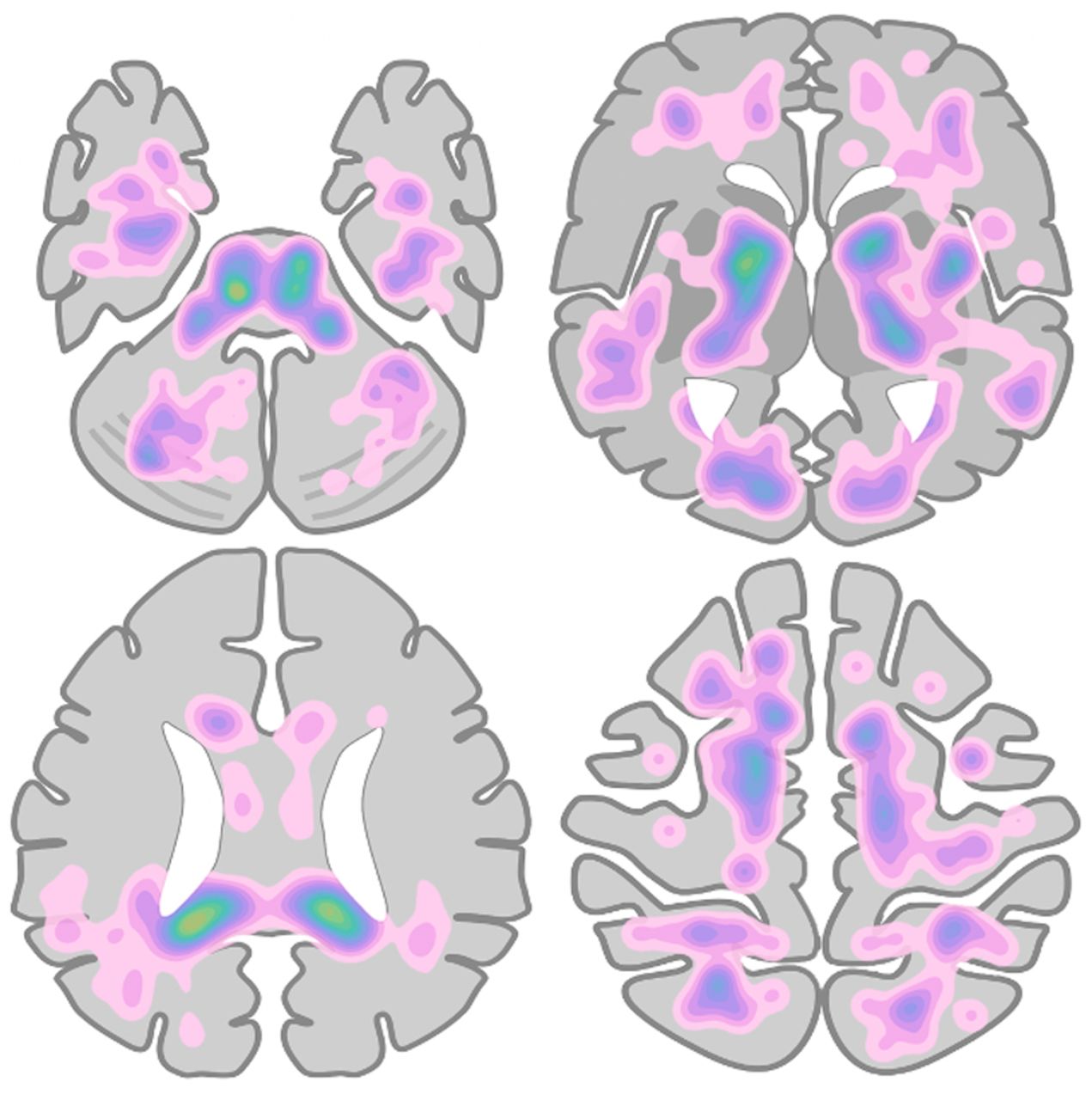
- Figure 2
Heatmap of microhaemorrhage density and distribution across all patients measured, based on manual labelling of microhaemorrhage locations on a simplified brain schematic.


Tables
- Table 1
Microhaemorrhage score and additional imaging findings for each patient (1–10)
1 2 3 4 5 6 7 8 9 10 Median Brainstem 5 5 5 7 0 1 – 0 4 0 4.00 Cerebellum 1 2 2 19 4 3 – 0 0 2 2.00 Middle cerebellar peduncle 0 4 3 7 0 0 – 0 0 0 0.00 Basal ganglia 0 2 3 5 2 0 – 0 1 1 1.00 Thalamus 0 0 0 1 0 0 – 0 0 0 0.00 Internal capsule 1 2 0 19 2 2 – 0 6 0 2.00 External capsule 0 9 0 7 0 0 – 0 2 0 0.00 Corpus callosum 17 2 12 52 5 4 – 1 15 14 12.00 Genu 5 16 1 7 1 0 – 0 7 2 2.00 Body 0 2 1 8 0 1 – 1 0 0 1.00 Splenium 12 4 10 37 4 3 – 0 8 12 8.00 Deep periventricular white matter 0 10 0 20 0 5 – 2 0 0 0.00 Frontal 0 0 0 51 0 16 – 2 4 3 2.00 Parietal 0 18 0 35 4 7 – 2 6 9 6.00 Temporal 0 8 0 30 3 2 – 9 1 3 3.00 Occipital 0 4 0 16 1 4 – 0 0 3 1.00 Insula 0 4 0 1 0 0 – 0 0 0 0.00 T2 signal change No Yes No Yes No No No No No No Restricted diffusion No No No No No No No No No No Macroscopic haemorrhage No No No Yes No No No No No No Total 24 70 25 270 21 44 – 16 39 35 35.00 Bold values refer to theareas with the greatest number of microhaemorrhages.
- Table 2
Summary of patient demographics and clinical features
1* 2 3 4 5 6 7 8 9 10 Median Age (years) 52 51 60 62 55 57 66 54 66 48 56.00 Sex Female Male Male Male Male Male Male Female Male Male – Premorbid condition Hypertension Yes Yes Yes – – Yes Yes – – – – Chronic kidney disease Yes – Yes – – – Yes – – – – Diabetes mellitus – – – – – Type 2 Type 2 Type 1 – Type 2 – Obesity – – – – Yes – Yes – – – – Hypercholesterolaemia – – – – – Yes Yes – – Yes – Respiratory
condition– – – – – – COPD – – – – Other – – Multiple myeloma Situs inversus – IHD – Polymyalgia – – ARDS Yes Yes Yes Yes Yes Yes Yes Yes Yes Yes – Reason for ITU admission T1RF T1RF T1RF T1RF T1RF T1RF T1RF T1RF T1RF T1RF – Length of
intubation (days)12 29 25 25 22 21 43 33 38 5 25.00 ECMO duration (days) – – – – – – – – – 14 0 Haemodialysis CVVHDF – CVVHDF – CVVHDF CVVHDF CVVHDF CVVHDF CVVHDF – – Heparinisation Yes Yes Yes Yes Yes Yes Yes Yes Yes Yes – Neurological presentation AMS AMS AMS Right-side weakness AMS AMS AMS Abnormal ventilation Tremors Seizures – Highest recorded blood pressure 190/70 150/112 110/80 – 143/96 208/69 163/74 172/81 137/77 142/91 – Biochemistry and haematology, worst value pH (7.35–7.45) – 7.12 7.15 7.05 7.05 7.09 7.19 6.95 7.13 7.15 7.12 PaCO2 (4.7–6 kPa) – 11.3 9.9 19.4 11.4 14.2 7.9 17.7 14.9 16.1 14.20 PaO2 (10.0–13.3 kPa) – 5.9 8.4 6.6 4.1 5.3 6.5 8.1 8.2 8.1 6.60 LDH (125–243 units/L) 609 345 774 410 1096 663 533 229 591 837 600.00 Hb (115–155 g/L) 72 68 68 71 68 73 63 64 66 91 68.00 WCC (4.2–10.6×10⁹/L) 14.4 19.9 31.6 20.4 29.9 14.6 19.3 25.6 24.3 31.3 22.35 Lymphocytes (1.1–3.6×10⁹/L) 2.2 0.4 0.6 0.3 0.6 0.5 0.2 0.3 0.3 0.7 0.45 Neutrophils (2.0–7.1×10⁹/L) 10.3 17.3 15.9 18.4 15.7 16.3 17.8 24.1 22.1 25.6 17.55 Platelets (highest/lowest)
(130–370×109/L)537/438 732/189 318/100 285/142 525/175 342/143 298/102 547/138 733/369 871/280 537/143 Urea (2.5–7.8) 44.3 14.9 51 20.3 41.3 49.8 42.6 26.9 43.3 7.7 41.95 CRP (<5 mg/L) 49 426 342 349 401 365 220 284 266 175 313.00 Ferritin (20–300 ng/mL) 2714 3607 3762 7569 4003 739 710 526 4916 12 592 3684.50 PT (12.8–17.4 s) 15.6 18.5 20 17.5 14.9 15.5 19.8 18.2 16.2 15.3 16.85 Fibrinogen
(1.9–4.3 g/L)5.1 8.6 9.6 8.7 4.3 10.3 7.5 8.6 7.9 7.5 8.25 D-dimer (<500 ng/mL) 2010 20 000 20 000 7569 6309 20 000 12 064 5569 13 148 7060 9816.50 Haematocrit (0.39%–0.50%) 0.224 0.424 0.44 0.3 0.41 0.4 0.36 0.34 0.289 0.41 0.38 DIC score 3 4 4 3 4 4 4 4 4 4 4.00 Systemic thrombosis No No No No No No IJV thrombus No DVT No Extent of consolidation on thoracic imaging – Severe Severe Severe Severe Severe Severe Severe Severe Severe MRI head day from admission 24 38 37 32 29 24 53 59 58 41 37.50 MARS 24 70 25 270 21 44 – 16 39 35 60.44 Bold values highlight the areas with the greatest median number of microhaemorrhages.
*Patient 1 has incomplete biochemistry information due to their initial care being at an external institution.
AMS, altered mental state; ARDS, acute respiratory distress syndrome; COPD, chronic obstructive pulmonary disease; CRP, c-reactive protein; CVVHDF, continuous venovenous haemodiafiltration; DIC, disseminated intravascular coagulation; ECMO, extracorporeal membrane oxygenation; Hb, haemoglobin; IHD, ischaemic heart disease; IJV, internal jugular vein; ITU, intensive therapy unit; LDH, lactate dehydrogenase; MARS, microbleed anatomical scale; Pa02, partial pressure of oxygen; PaC02, partial pressure of carbon dioxide; PT, prothrombin time; T1RF, type 1 respiratory failure; WCC, white cell count.


