

Article Figures & Data
Figures
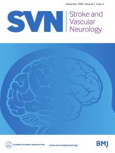
- Figure 1
Flow chart of the process used to select subjects. AIS, acute ischaemic stroke; END, early neurological deterioration; ILASO, intracranial large artery stenosis or occlusion; IVT, intravenous thrombolysis; SWI, susceptibility-weighted imaging; PH, parenchymatous haemorrhage.
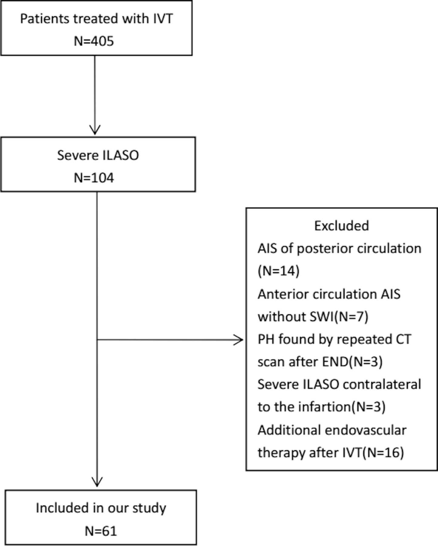
- Figure 2
(1) The case of a 74-year-old woman who suffered from AIS for 2 hours. In the left haemisphere, MHVs in M1 (A), M4 and M5 (B) were evident on SWI, and defined as subtle MHVs. (2) The case of a 68-year-old woman who suffered from AIS for 3 hours. In the left haemisphere, MHVs in M1, M2, M3 (C) and M4, M5, M6 (D) were evident on SWI, and defined as moderate MHVs. (3) The case of a 42-year-old man who suffered from AIS for 2 hours. In the left haemisphere, MHVs in M1, M2, M3 (E) and M4, M5, M6, Deep (F) were evident on SWI, and defined as extensive MHVs. AIS, acute ischaemic stroke; Deep, deep white matter; MHVs, multiple hypointense vessels; M1, anterior MCA cortex; M2, MCA cortex lateral to the insular cortex; M3, posterior MCA cortex; M4, M5, M6, the anterior, lateral and posterior MCA territories immediately superior to M1, M2 and M3; MCA, middle cerebral artery; SWI, susceptibility-weighted imaging.


Tables
- Table 1
The parameters for each sequence of MRI scan
TR
(ms)TE
(ms)FOV
(mm2/mm3)ST/gap
(mm)TA
(s)ETL ESP
(ms)Slice no Scanning time (s) T1WI 1500 11 220×185 4/1.2 86 27 11 25 86 T2WI 4720 96 220×199 4/1.2 110 11 12 25 110 FLAIR 9000 84 230×187 5/1.5 110 5 10.5 25 110 DWI 4640 67 230×218 4/1.2 104 – – 25 104 SWI 27 20 240×195 3/0.6 88 – – Volume scan 143 3D-TOF-MRA 21 3.42 200×160×160 0.7/–0.14 216 – – Volume scan 216 3D-TOF-MRA, three-dimensional time-of-flight MR angiography; DWI, diffusion-weighted imaging; ESP, echo spacing; ETL, echo train length; FLAIR, fluid-attenuated inversion recovery; FOV, field of view; ST, slice thickness; SWI, Susceptibility-weighted imaging; TA, time of acquisition; TE, time of echo; TR, time of repetition; T1WI, T1-weighted imaging; T2WI, T2-weighted imaging.
- Table 2
Demographic and clinical characteristics of the study cohort
Characteristics Mean(SD)/median
(IQR)/n (%) n=61Age (years)* 62.4±12.6 Men (n, %) 51 (83.6) Hypertension (n, %) 37 (60.7) Diabetes mellitus (n, %) 15 (24.6) Smokers/ex-smokers (n, %) 28 (45.9) Atrial fibrillation (n, %) 15 (24.6) Previous stroke (n, %) 11 (18.0) OTT (minutes)* 203.5±54.1 NIHSS score on admission† 9 (5–14) SBP on admission (mm Hg)* 150.6±22.8 DBP on admission (mm Hg)* 89.3±16.9 NLR† 2.4 (1.7–5.9) BG level on admission(mmol/L)* 7.0±2.3 Homocysteine (mmol/L)* 14.4±6.9 END (n, %) 20 (32.8) Severe MCA stenosis (n, %) 31 (50.8) MCA occlusion (n, %) 17 (27.9) Severe ICA stenosis (n, %) 4 (6.5) ICA occlusion (n, %) 9 (14.8) DWI-ASPECT score† 7 (5–8) MHVs (n, %) 35 (57.4) Subtle (n, %) 8 (13.1) Moderate (n, %) 23 (37.7) Extensive (n, %) 4 (6.6) SVD burden† 0 (0–1) *Mean±SD.
†Median (IQR).
ASPECT, Alberta Stroke Programme Early CT ; BG, blood glucose; DBP, diastolic blood pressure; DWI, diffusion-weighted imaging; END, early neurological deterioration; ICA, internal carotid artery; MCA, middle cerebral artery; MHVs, multiple hypointense vessels; NIHSS, National Institutes of Health Stroke Scale; NLR, neutrophil to lymphocyte ratio; OTT, onset to treatment time; SBP, systolic blood pressure; SVD, small vessel disease.
- Table 3
Risk factors of END determined by univariable analysis
Variable END P value With (n=20) Without (n=41) Age* (year) 63.0 (11.1) 62.2 (13.4) 0.818 Men† (n, %) 18 (90.0) 33 (80.5) 0.346 Hypertension† (n, %) 15 (75.0) 22 (53.7) 0.109 Diabetes† (n, %) 6 (30.0) 9 (22.0) 0.493 Smokers/ex-smokers† (n, %) 10 (50.0) 18 (43.9) 0.654 Atrial fibrillation‡ (n, %) 5 (25.0) 10 (24.4) 1.000 Previous stroke† (n, %) 5 (25.0) 6 (14.6) 0.323 OTT* (minutes) 221.6 (46.8) 194.7 (55.9) 0.048 NIHSS score on admission§ 7.5 (5–11) 10 (5.5–15.5) 0.100 SBP on admission*
(mm Hg)151.8 (19.5) 150.0 (24.5) 0.769 DBP on admission*
(mm Hg)92.6 (17.4) 87.7 (16.7) 0.301 NLR§ 2.7 (1.7–7.5) 2.4 (1.7–4.5) 0.484 BG on admission* (mmol/L) 7.3 (2.7) 6.3 (1.1) 0.041 Homocysteine* (mmol/L) 15.7 (9.1) 13.8 (5.5) 0.305 DWI-ASPECT score§ 6.0 (5.0–7.5) 7.0 (5.5–8.0) 0.211 MHVs† (n, %) 0.003 None 4 (20.0) 22 (53.7) Subtle 2 (10.0) 6 (14.6) Moderate 11 (55.0) 12 (29.3) Extensive‡ 3 (15.0) 1 (2.4) SVD‡ 0 (0–1) 0 (0–2) 0.747 *Mean (SD), t-test.
†n(%), X2 test.
‡Fisher’s exact test.
§Median (IQR), Mann-Whitney U test.
ASPECT, Alberta Stroke Programme Early CT Score; BG, blood glucose; DBP, diastolic blood pressure; DWI, diffusion-weighted imaging; END, early neurological deterioration; MHVs, multiple hypointense vessels; NIHSS, National Institutes of Health Stroke Scale; NLR, neutrophil to lymphocyte ratio; OTT, onset to treatment time; SBP, systolic blood pressure; SVD, small vessel burden.
- Table 4
The identification of predictors for END by multivariate logistic regression
Variable END Unadjusted p value Adjusted OR (95% CI) Adjusted p value Crude OR (95% CI) Age 1.005 (0.963 to 1.049) 0.814 1.031 (0.973 to 1.093) 0.306 Sex 0.395 (0.077 to 2.031) 0.266 0.303 (0.042 to 2.194) 0.237 OTT 1.010 (0.999 to 1.021) 0.073 1.011 (0.998 to 1.023) 0.090 NIHSS 0.902 (0.805 to 1.010) 0.073 0.913 (0.805 to 1.035) 0.154 BG on admission 0.759 (0.531 to 1.084) 0.129 1.012 (0.999 to 1.026) 0.067 MHVs – 0.043 – 0.049 None (as reference) – – – – Subtle 1.833 (0.268 to 12.536) 0.537 2.083 (0.287 to 15.111) 0.468 Moderate 5.042 (1.316 to 19.317) 0.018 5.446 (1.360 to 21.800) 0.017 Extensive 16.500 (1.353 to 201.290) 0.028 15.240 (1.200 to 193.544) 0.036 BG, blood glucose; END, early neurological deterioration; MHVs, Multiple hypointense vessels; NIHSS, National Institutes of Health Stroke Scale; OTT, onset to treatment time.
Supplementary data


