

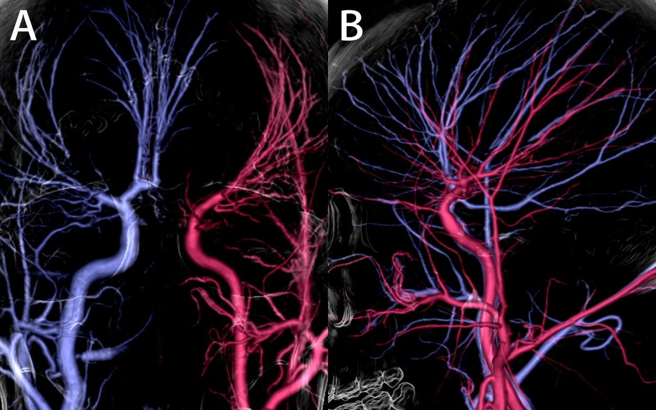
A 45-year-old man diagnosed with multisystemic smooth muscle dysfunction syndrome (MSMDS) was referred for the evaluation of recurrent dizziness that had persisted for 2 years. MRI revealed a right frontal lobe cerebral infarction, enlargement of the bilateral ventricular trigones and hypoplasia of the corpus callosum (figure 1A,B). MRA showed occlusion of the right middle cerebral artery (figure 1C,D). Cinematic rendering of CTA highlighted characteristic cerebrovascular manifestations of MSMDS: dilatation of the internal carotid artery from the cavernous to the clinoid segments, stenosis and occlusion of the distal intracranial circulation and an abnormally straight course of intracranial arteries (figure 2, online supplemental video S1).1 Cinematic rendering of CTA provides highly realistic 3D reconstructed images, enabling multi-angle and multi-plane rotational visualisation of vascular structures. This technique offers significant educational value, aiding neurointerventionalists in preoperative planning and improving communication between neurosurgeons and patients.2 There are currently no standard indications for neurosurgical revascularisation for MSMDS, which may be due in part to the paucity of reported cases and the high risk of postoperative stroke in the limited number of cases.1 3 Nevertheless, given the progressive nature of MSMDS and the risk of recurrent strokes, early surgical intervention may be considered in selected cases to prevent further neurological deterioration. MSMDS is an autosomal dominant condition caused by a mutation in the ACTA2 gene.4 Neurosurgeons should suspect MSMDS in patients presenting with recurrent strokes and characteristic cerebrovascular findings, and early genetic investigations should be prioritised to confirm the diagnosis.5
Supplementary video

MRI and MRA imaging. (A) Diffusion-weighted image showed right frontal lobe cerebral infarction (red arrow) and trigone of bilateral ventricles enlargement (white arrow). (B) T1 axial section showed hypoplasia of the corpus callosum. (C) Coronal and (D) sagittal MRI showed occlusion of the M1 segment of the right middle cerebral artery.

Cinematic rendering of CTA images of cerebral angiography in orthostatic (A) and lateral (B) positions. Cinematic rendering of CTA clearly depicts the anatomic contours of the bilateral internal carotid arteries; the photorealistic images produced by cinematic rendering could help neurosurgeons better familiarise themselves with this rare disease and its characteristic cerebrovascular manifestations.
Ethics statements
Patient consent for publication
Ethics approval
This study involves human participants and was approved by the ethics committee of Beijing Tiantan Hospital, and the approval number given by the ethical board was KY 2022-174-02. Participants gave informed consent to participate in the study before taking part.
Footnotes
GL and ML contributed equally.
Contributors LD drafted and edited the manuscript. ML provided funding and designed the study. GL and ML revised the article. All authors have read and approved the final manuscript. ML is the guarantor.
Funding This study was funded by the National Natural Science Foundation of China (82271319)
Competing interests None declared.
Provenance and peer review Not commissioned; externally peer reviewed.
Supplemental material This content has been supplied by the author(s). It has not been vetted by BMJ Publishing Group Limited (BMJ) and may not have been peer-reviewed. Any opinions or recommendations discussed are solely those of the author(s) and are not endorsed by BMJ. BMJ disclaims all liability and responsibility arising from any reliance placed on the content. Where the content includes any translated material, BMJ does not warrant the accuracy and reliability of the translations (including but not limited to local regulations, clinical guidelines, terminology, drug names and drug dosages), and is not responsible for any error and/or omissions arising from translation and adaptation or otherwise.
This is an open access article distributed in accordance with the Creative Commons Attribution Non Commercial (CC BY-NC 4.0) license, which permits others to distribute, remix, adapt, build upon this work non-commercially, and license their derivative works on different terms, provided the original work is properly cited, appropriate credit is given, any changes made indicated, and the use is non-commercial. See: http://creativecommons.org/licenses/by-nc/4.0/.


