
Main menu

  • Online first
    • Online first
  • Current issue
    • Current issue
  • Archive
    • Archive
  • Submit a paper
    • Online submission site
    • Instructions for authors
  • About the journal
    • About the journal
    • Editorial board
    • Instructions for authors
    • FAQs
    • Chinese Stroke Association
  • Help
    • Contact us
    • Feedback form
    • Reprints
    • Permissions
    • Advertising
  • BMJ Journals

User menu

  • Login

Search

  • Advanced search
  • BMJ Journals
  • Login
  • Facebook
  • Twitter
Stroke and Vascular Neurology

Advanced Search

  • Online first
    • Online first
  • Current issue
    • Current issue
  • Archive
    • Archive
  • Submit a paper
    • Online submission site
    • Instructions for authors
  • About the journal
    • About the journal
    • Editorial board
    • Instructions for authors
    • FAQs
    • Chinese Stroke Association
  • Help
    • Contact us
    • Feedback form
    • Reprints
    • Permissions
    • Advertising
Open Access

M2 middle cerebral artery dissection on 7T MRI

Xuewei Xie, Zhe Zhang, Qingle Kong, Hui Qu, Jing Jing
DOI: 10.1136/svn-2022-001557 Published 20 December 2022
Xuewei Xie
1 Department of Neurology, Beijing Tiantan Hospital, Capital Medical University, Beijing, China
2 China National Clinical Research Center for Neurological Diseases, Beijing, China
  • Find this author on Google Scholar
  • Find this author on PubMed
  • Search for this author on this site
  • ORCID record for Xuewei Xie
Zhe Zhang
2 China National Clinical Research Center for Neurological Diseases, Beijing, China
3 Tiantan Neuroimaging Center of Excellence, Beijing, China
  • Find this author on Google Scholar
  • Find this author on PubMed
  • Search for this author on this site
Qingle Kong
4 MR Collaboration, Siemens Healthineers Ltd, Beijing, China
  • Find this author on Google Scholar
  • Find this author on PubMed
  • Search for this author on this site
Hui Qu
1 Department of Neurology, Beijing Tiantan Hospital, Capital Medical University, Beijing, China
2 China National Clinical Research Center for Neurological Diseases, Beijing, China
  • Find this author on Google Scholar
  • Find this author on PubMed
  • Search for this author on this site
Jing Jing
1 Department of Neurology, Beijing Tiantan Hospital, Capital Medical University, Beijing, China
2 China National Clinical Research Center for Neurological Diseases, Beijing, China
3 Tiantan Neuroimaging Center of Excellence, Beijing, China
  • Find this author on Google Scholar
  • Find this author on PubMed
  • Search for this author on this site
  • Article
  • Figures & Data
  • eLetters
  • Info & Metrics
  • PDF
Loading

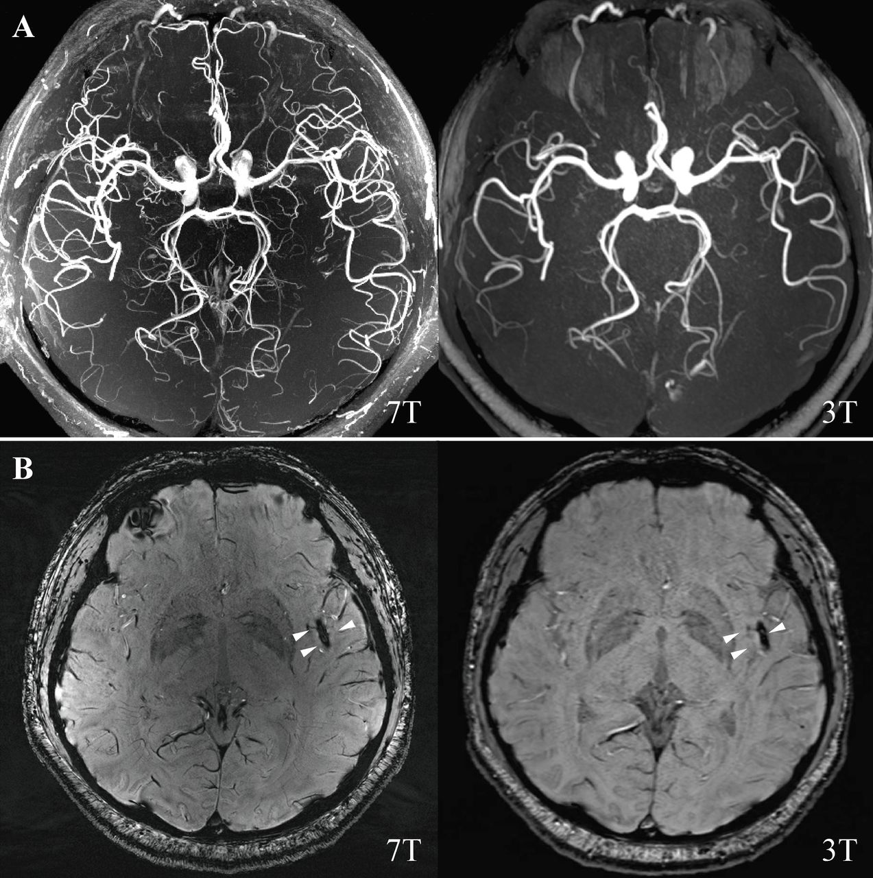
A 36-year-old man was admitted for acute right extremity weakness with multiple acute infarctions in the left middle cerebral artery (MCA) territory. 7T and 3T MRI scans were completed at 9 and 10 days after symptom onset (figure 1). There was susceptibility vessel sign, which represents red blood cell dominant intraluminal thrombus,1 2 at left distal M2 segment MCA on susceptibility-weighted imaging (figure 1B). 7T vessel wall MRI showed a typical feature of a long segment dissection at the left distal M2 segment of MCA with a mural haematoma and the residual lumen (figure 2A), which was not seen on 3T (figure 2B). Vessel wall MRI was a valuable tool for the diagnosis of intracranial artery dissection.2 3 This case indicated that 7T vessel wall MRI may provide better detail on the pathological change than traditional 3T MRI for diagnosing distal intracranial arterial dissection, the diagnose otherwise could be missed.

Figure 1
  • Download figure
  • Open in new tab
  • Download powerpoint
Figure 1

(A) 7T and 3T magnetic resonance angiography. (B) 7T and 3T SWI were showing blooming hypointense signals (susceptibility vessel sign) in the distal M2 segment of the left MCA (white triangle). MCA, middle cerebral artery; SWI, susceptibility-weighted imaging.

Figure 2
  • Download figure
  • Open in new tab
  • Download powerpoint
Figure 2

(A) 7T 3D T1-SPACE MRI (MAGNETOM Terra, Siemens Healthcare, Erlangen, Germany. Voxel size=0.4×0.4×0.4 mm3) showing left distant M2 segment MCA mural haematoma and residual lumen, and axial reformatted images showing a semilunar hyperintense signal caused by a mural haematoma (yellow triangle) and an eccentric hypointense signal attributable to the residual lumen (white triangle). (B) 3T 3D T1-SPACE MRI (MAGNETOM Prisma, Siemens Healthcare, Erlangen, Germany. Voxel size=0.54×0.54×0.54 mm3) showing left distant M2 segment MCA intraluminal thrombus formation and axial reformatted images showing only hyperintense signal (white triangle) with an unclear residual lumen. MCA, middle cerebral artery.

Ethics statements

Patient consent for publication

Consent obtained directly from patient(s)

Footnotes

  • XX and ZZ contributed equally.

  • HQ and JJ contributed equally.

  • Contributors XX: drafting of the manuscript. ZZ: analysis and interpretation of data. QK: analysis and interpretation of data. HQ: acquisition of images and data. JJ: acquisition of images and data, and drafting of the manuscript.

  • Funding The authors have not declared a specific grant for this research from any funding agency in the public, commercial or not-for-profit sectors.

  • Competing interests None declared.

  • Provenance and peer review Not commissioned; externally peer reviewed.

https://creativecommons.org/licenses/by/4.0/

This is an open access article distributed in accordance with the Creative Commons Attribution 4.0 Unported (CC BY 4.0) license, which permits others to copy, redistribute, remix, transform and build upon this work for any purpose, provided the original work is properly cited, a link to the licence is given, and indication of whether changes were made. See: https://creativecommons.org/licenses/by/4.0/.

References

  1. ↵
    1. Liebeskind DS ,
    2. Sanossian N ,
    3. Yong WH , et al
    . CT and MRI early vessel signs reflect clot composition in acute stroke. Stroke 2011;42:1237–43.doi:10.1161/STROKEAHA.110.605576 pmid:http://www.ncbi.nlm.nih.gov/pubmed/21393591
    OpenUrlAbstract/FREE Full Text
  2. ↵
    1. Wu Y ,
    2. Wu F ,
    3. Liu Y , et al
    . High-Resolution magnetic resonance imaging of Cervicocranial artery dissection: imaging features associated with stroke. Stroke 2019;50:3101–7.doi:10.1161/STROKEAHA.119.026362 pmid:http://www.ncbi.nlm.nih.gov/pubmed/31514693
    OpenUrlPubMed
  3. ↵
    1. Chen H ,
    2. Li Z ,
    3. Luo B , et al
    . Anterior cerebral artery dissection diagnosed using high-resolution MRI. Neurology 2015;85:481.doi:10.1212/WNL.0000000000001813 pmid:http://www.ncbi.nlm.nih.gov/pubmed/26238819
    OpenUrlPubMed
PreviousNext
Back to top
Vol 7 Issue 6 Table of Contents
Stroke and Vascular Neurology: 7 (6)
  • Table of Contents
  • Table of Contents (PDF)
  • About the Cover
  • Index by author
  • Front Matter (PDF)
Email

Thank you for your interest in spreading the word on Stroke and Vascular Neurology.

NOTE: We only request your email address so that the person you are recommending the page to knows that you wanted them to see it, and that it is not junk mail. We do not capture any email address.

Enter multiple addresses on separate lines or separate them with commas.
M2 middle cerebral artery dissection on 7T MRI
(Your Name) has sent you a message from Stroke and Vascular Neurology
(Your Name) thought you would like to see the Stroke and Vascular Neurology web site.
CAPTCHA
This question is for testing whether or not you are a human visitor and to prevent automated spam submissions.
Print
Alerts
Sign In to Email Alerts with your Email Address
Citation Tools
M2 middle cerebral artery dissection on 7T MRI
Xuewei Xie, Zhe Zhang, Qingle Kong, Hui Qu, Jing Jing
Stroke and Vascular Neurology Dec 2022, 7 (6) 550; DOI: 10.1136/svn-2022-001557

Citation Manager Formats

  • BibTeX
  • Bookends
  • EasyBib
  • EndNote (tagged)
  • EndNote 8 (xml)
  • Medlars
  • Mendeley
  • Papers
  • RefWorks Tagged
  • Ref Manager
  • RIS
  • Zotero
Cite This
  • APA
  • Chicago
  • Endnote
  • MLA
Loading
M2 middle cerebral artery dissection on 7T MRI
Xuewei Xie, Zhe Zhang, Qingle Kong, Hui Qu, Jing Jing
Stroke and Vascular Neurology Dec 2022, 7 (6) 550; DOI: 10.1136/svn-2022-001557
Download PDF

Share
M2 middle cerebral artery dissection on 7T MRI
Xuewei Xie, Zhe Zhang, Qingle Kong, Hui Qu, Jing Jing
Stroke and Vascular Neurology Dec 2022, 7 (6) 550; DOI: 10.1136/svn-2022-001557
Reddit logo Twitter logo Facebook logo Mendeley logo
Respond to this article
  • Tweet Widget
  • Facebook Like
  • Google Plus One
  • Article
    • Ethics statements
    • Footnotes
    • References
  • Figures & Data
  • eLetters
  • Info & Metrics
  • PDF

Related Articles

Cited By...

More in this TOC Section

  • Cerebrovascular cinematic rendering of multisystemic smooth muscle dysfunction syndrome
  • Pictures of stroke and cerebral vasculature: novel insights and detailed visualisation of U-shaped elastin distribution and disorganised fibre arrangement at carotid bifurcations by advanced techniques
Show more Pictures of stroke and cerebral vasculature

Similar Articles


CONTENT

  • Latest content
  • Current issue
  • Archive
  • eLetters
  • Sign up for email alerts
  • RSS

JOURNAL

  • About the journal
  • Editorial board
  • Recommend to librarian
  • Chinese Stroke Association

AUTHORS

  • Instructions for authors
  • Submit a paper
  • Track your article
  • Open Access at BMJ

HELP

  • Contact us
  • Reprints
  • Permissions
  • Advertising
  • Feedback form

© 2025 Chinese Stroke Association