
Main menu

  • Online first
    • Online first
  • Current issue
    • Current issue
  • Archive
    • Archive
  • Submit a paper
    • Online submission site
    • Instructions for authors
  • About the journal
    • About the journal
    • Editorial board
    • Instructions for authors
    • FAQs
    • Chinese Stroke Association
  • Help
    • Contact us
    • Feedback form
    • Reprints
    • Permissions
    • Advertising
  • BMJ Journals

User menu

  • Login

Search

  • Advanced search
  • BMJ Journals
  • Login
  • Facebook
  • Twitter
Stroke and Vascular Neurology

Advanced Search

  • Online first
    • Online first
  • Current issue
    • Current issue
  • Archive
    • Archive
  • Submit a paper
    • Online submission site
    • Instructions for authors
  • About the journal
    • About the journal
    • Editorial board
    • Instructions for authors
    • FAQs
    • Chinese Stroke Association
  • Help
    • Contact us
    • Feedback form
    • Reprints
    • Permissions
    • Advertising
Open Access

Cellular and molecular imaging for stem cell tracking in neurological diseases

Xiaoxia Yang, De-Cai Tian, Wenyan He, Wei Lv, Junwan Fan, Haowen Li, Wei-Na Jin, Xia Meng
DOI: 10.1136/svn-2020-000408 Published 29 October 2020
Xiaoxia Yang
1China National Clinical Research Center for Neurological Diseases, Capital Medical University, Beijing Tiantan Hospital, Beijing, China
2Department of Neurology, Tianjin Neurological Institute, Tianjin Medical University General Hospital, Tianjin, China
  • Find this author on Google Scholar
  • Find this author on PubMed
  • Search for this author on this site
De-Cai Tian
1China National Clinical Research Center for Neurological Diseases, Capital Medical University, Beijing Tiantan Hospital, Beijing, China
  • Find this author on Google Scholar
  • Find this author on PubMed
  • Search for this author on this site
  • ORCID record for De-Cai Tian
Wenyan He
1China National Clinical Research Center for Neurological Diseases, Capital Medical University, Beijing Tiantan Hospital, Beijing, China
  • Find this author on Google Scholar
  • Find this author on PubMed
  • Search for this author on this site
Wei Lv
1China National Clinical Research Center for Neurological Diseases, Capital Medical University, Beijing Tiantan Hospital, Beijing, China
  • Find this author on Google Scholar
  • Find this author on PubMed
  • Search for this author on this site
Junwan Fan
1China National Clinical Research Center for Neurological Diseases, Capital Medical University, Beijing Tiantan Hospital, Beijing, China
  • Find this author on Google Scholar
  • Find this author on PubMed
  • Search for this author on this site
Haowen Li
1China National Clinical Research Center for Neurological Diseases, Capital Medical University, Beijing Tiantan Hospital, Beijing, China
  • Find this author on Google Scholar
  • Find this author on PubMed
  • Search for this author on this site
Wei-Na Jin
1China National Clinical Research Center for Neurological Diseases, Capital Medical University, Beijing Tiantan Hospital, Beijing, China
  • Find this author on Google Scholar
  • Find this author on PubMed
  • Search for this author on this site
Xia Meng
1China National Clinical Research Center for Neurological Diseases, Capital Medical University, Beijing Tiantan Hospital, Beijing, China
  • Find this author on Google Scholar
  • Find this author on PubMed
  • Search for this author on this site
  • ORCID record for Xia Meng
  • Article
  • Figures & Data
  • eLetters
  • Info & Metrics
  • PDF
Loading

Article Figures & Data

Figures

  • Tables
  • Figure 1
    • Download figure
    • Open in new tab
    • Download powerpoint
    Figure 1

    Timeline for HPF labelling MSCs. When the cultured MSCs grow to 80%–90% density, heparin, protamine and ferulic acid are added in sequence and mixed for 5 min. After 2–4 hours of cocultivation, 10%–20% FBS was added and the culture was continued for 24 hours. After washing with PBS and heparin, the culture was continued for 15 min to obtain HPF nanocomplex. HPF, ferumoxytol–heparin–protamine; MSC, mesenchymalstem cell;FBS, fatal bovine serun; PBS, phosphate buffered solution.
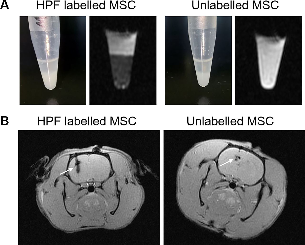
  • Figure 2
    • Download figure
    • Open in new tab
    • Download powerpoint
    Figure 2

    HPF nanocomplex combined with MRI can track and monitor MSCs. (A). The images show HPF-labelled and unlabelled MscS in the tube in vitro by the T2*-weighted MRI. (B.) the images show HPF-labelled and unlabelled MscS in the brain striatum of rats on the third day after stereotactic injection by the T2*-weighted MRI. HPF, ferumoxytol–heparin–protamine; MSC, mesenchymalstem cell.

Tables

  • Figures
  • Table 1

    Labelling and tracking stem cells in neurological diseases

    DiseaseCell typeTracking technologyTracerSpeciesReferences
    StrokeMSCMRIMolday ION-Rhodamine BPigs/dogs/ RatsWalczak et al, 201751
    StrokeMSCMRI/NIRFBCN-dual-NPsMiceLim et al, 201952
    StrokeMSCMRI/PAIGRMNBsMiceChen et al, 201553
    StrokeNSCMRI/FLILV-FTH-EGFPMiceZhang et al, 201754
    StrokeMSCMRI/SPECT/CT125I-fSiO4@SPIOsRatsTang et al, 201555
    PDNSCMRIMNPRatsGomez et al, 201510
    ADMSCMRIFerumoxytolMiceLee et al, 201767
    ALShGRPMRIPFCMiceRichard et al, 201972
    TBIiPS cellMRISPIORatsJiang et al, 201978
    SCIBM-MSCMRIGd-DTPA-FARatsZhang et al, 201786
    • AD, Alzheimer’s disease; ALS, amyotrophic lateral sclerosis; BCN-dual-NPs, bicyclic nonyne conjugated ethylene glycol chitosan nanoparticles; BM-MSC, bone marrow mesenchymal stem cell; iPS cell, induced pluripotent stem cell; FLI, fluorescence imaging; Gd-DTPA-FA, gadolinium-dimethylene penta-acetic acid-containing nanoparticles; GRMNBs, multi-gold nanorods crystal-seeded magnetic mesoporous silica nanobeads; hGRP, human glial-restricted progenitor; 125I-fSiO4@SPIOs, fluorescent silica-coated SPIOs with 125iodine; LV-FTH-EGFP, a lentiviral vector encoding ferritin heavy chain (FTH) and enhanced green fluorescent protein (EGFP); MNP, magnetic nanoparticle; MSC, mesenchymal stem cells; NIRF, near-infrared fluorescent; NSC, neural stem cell; PAI, photoacoustic imaging; PD, Parkinson’s disease; PET, positron emission tomography; PFC, perfluorocarbon; SCI, spinal cord injury; SPECT, single-photon emission CT; SPIO, superparamagnetic iron oxide; TBI, traumatic brain injury.

PreviousNext
Back to top
Vol 10 Issue 1 Table of Contents
Stroke and Vascular Neurology: 10 (1)
  • Table of Contents
  • Table of Contents (PDF)
  • About the Cover
  • Index by author
  • Ed Board (PDF)
Email

Thank you for your interest in spreading the word on Stroke and Vascular Neurology.

NOTE: We only request your email address so that the person you are recommending the page to knows that you wanted them to see it, and that it is not junk mail. We do not capture any email address.

Enter multiple addresses on separate lines or separate them with commas.
Cellular and molecular imaging for stem cell tracking in neurological diseases
(Your Name) has sent you a message from Stroke and Vascular Neurology
(Your Name) thought you would like to see the Stroke and Vascular Neurology web site.
CAPTCHA
This question is for testing whether or not you are a human visitor and to prevent automated spam submissions.
Print
Alerts
Sign In to Email Alerts with your Email Address
Citation Tools
Cellular and molecular imaging for stem cell tracking in neurological diseases
Xiaoxia Yang, De-Cai Tian, Wenyan He, Wei Lv, Junwan Fan, Haowen Li, Wei-Na Jin, Xia Meng
Stroke and Vascular Neurology Oct 2020, svn-2020-000408; DOI: 10.1136/svn-2020-000408

Citation Manager Formats

  • BibTeX
  • Bookends
  • EasyBib
  • EndNote (tagged)
  • EndNote 8 (xml)
  • Medlars
  • Mendeley
  • Papers
  • RefWorks Tagged
  • Ref Manager
  • RIS
  • Zotero
Cite This
  • APA
  • Chicago
  • Endnote
  • MLA
Loading
Cellular and molecular imaging for stem cell tracking in neurological diseases
Xiaoxia Yang, De-Cai Tian, Wenyan He, Wei Lv, Junwan Fan, Haowen Li, Wei-Na Jin, Xia Meng
Stroke and Vascular Neurology Oct 2020, svn-2020-000408; DOI: 10.1136/svn-2020-000408
Download PDF

Share
Cellular and molecular imaging for stem cell tracking in neurological diseases
Xiaoxia Yang, De-Cai Tian, Wenyan He, Wei Lv, Junwan Fan, Haowen Li, Wei-Na Jin, Xia Meng
Stroke and Vascular Neurology Oct 2020, svn-2020-000408; DOI: 10.1136/svn-2020-000408
Reddit logo Twitter logo Facebook logo Mendeley logo
  • Tweet Widget
  • Facebook Like
  • Google Plus One
  • Article
    • Abstract
    • Introduction
    • Techniques for labelling and tracking SCs
    • SC tracing in an animal model and clinical trials of neurological diseases
    • Conclusion
    • Footnotes
    • References
  • Figures & Data
  • eLetters
  • Info & Metrics
  • PDF

Related Articles

Cited By...

More in this TOC Section

  • Design of trials in lacunar stroke and cerebral small vessel disease: review and experience with the LACunar Intervention Trial 2 (LACI-2)
  • Anti-stroke biologics: from recombinant proteins to stem cells and organoids
  • Central post-stroke pain: advances in clinical and preclinical research
Show more Review

Similar Articles


CONTENT

  • Latest content
  • Current issue
  • Archive
  • eLetters
  • Sign up for email alerts
  • RSS

JOURNAL

  • About the journal
  • Editorial board
  • Recommend to librarian
  • Chinese Stroke Association

AUTHORS

  • Instructions for authors
  • Submit a paper
  • Track your article
  • Open Access at BMJ

HELP

  • Contact us
  • Reprints
  • Permissions
  • Advertising
  • Feedback form

© 2025 Chinese Stroke Association