

Article Figures & Data
Figures
- Figure 1
Illustration of VWF mechanoactivation (inset) and the step-by-step process of GPIb-mediated platelet mechanosensing. VWF is activated by shear flow in two steps: it is first globally elongated from a globular to an extended conformation, followed by the relief of its A1 domain autoinhibition that enables binding to platelet GPIbα. Once VWF is bound to a platelet, force from the shear flow transmits from VWF to GPIbα and triggers a series of GPIbα conformational changes and allosteric effects. These events would result in the reinforcement of VWF–GPIb interaction as well as the initiation of a mechanosignaling pathway that eventually leads to intracellular calcium release and GPIIb/IIIa activation. Note: platelet GPVI and its interaction with collagen are also depicted in this graph. Agents and drugs that have the potential to inhibit arterial thrombosis by targeting GPIb-mediated or GPVI-mediated platelet binding and mechanosensing, and their respective targets, are indicated, corresponding to table 1.
- Figure 2
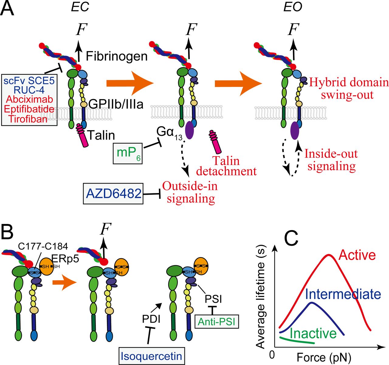
GPIIb/IIIa mediated platelet mechanosensing. (A) The extended-close (EC) GPIIb/IIIa receives mechanosignals from its bound ligand and mediates outside-in signaling, which can subsequently upregulate GPIIb/IIIa themselves towards the active state with an extended-open (EO) conformation. During the process, the adaptor protein talin that is originally associated with the cytoplasmic tail of GPIIIa will detach and be replaced by a Gα13 molecule, which will be ensued by a second wave of talin attachment to in turn replace Gα13 (not depicted here). (B) Redox regulation of GPIIb/IIIa binding via ERp5, PSI and PDI. Force pulling on the GPIIb/IIIa headpiece via a bound Arg-Gly-Asp (RGD)-bearing ligand can facilitate ERp5 to reduce the C177–C184 disulfide bond in the βI domain, which in turn accelerates ligand dissociation from GPIIb/IIIa. PSI domain has endogenous thiol isomerase function, which reinforces GPIIb/IIIa binding. Extracellular PDI can also induce thiol-disulfide exchange in GPIIb/IIIa and enhances its binding capacity. (C) In the resting bent-closed (BC) state, GPIIb/IIIa binding to fibronectin manifests a slip bond (a small catch bond if against fibrinogen). Once upregulated to the intermediate extended-closed (EC) state, GPIIb/IIIa binding to both ligands will adopt a strong catch bond, which will become even stronger when the integrin is further activated to the EO state. Agents and drugs (green: preclinical phase; blue: undergoing clinical trials; red: FDA approved) that have the potential to inhibit arterial thrombosis by targeting GPIIb/IIIa mechanosensing, and their respective targets, are indicated, corresponding to table 1. ‘Anti-PSI’ represents antibodies PSI A1, PSI B1, PSI C1 and PSI E1. ERp5, endoplasmic reticulum 5; PDI, protein disulfide isomerase.


Tables
- Table 1
Novel antiplatelet agents targeting GPIb, GPIIb/IIIa and GPVI mechanosensing axes
Axis Target Agent Description Antithrombotic effect Phase of development Clinical trials gov identifier References GPIb VWF Lp (Q1238-E1260) The N-terminal polypeptide sequence of VWF-A1 that autoinhibits VWF-A1 binding to platelet GPIb. Inhibits platelet attachment on VWF in whole blood perfusion in vitro. Preclinical 57 14-3-3ζ MPαC Designed based on the 14-3-3ζ-binding sequence in GPIbα. Blocks 14-3-3ζ binding to GPIbα. Weakens GPIbα-VWF interaction and inhibits GPIbα mechanosignalling. Preclinical 92 93 VWF BAX 930 Recombinant ADAMTS13; cleaves VWF at A2 domain to weaken its activity. Inhibits arterial thrombosis. III NCT03393975 88 89 VWF NMC4 A monoclonal antibody against VWF-A1. Inhibits arterial thrombosis. Preclinical 73 VWF Caplacizumab A nanobody against VWF-A1. Showed inhibitory effects on thrombosis in primate models without causing adverse side effects. FDA approved 71 72 VWF ARC1779 An anti-VWF aptamer. Showed encouraging results in inhibiting thrombosis. II (terminated) NCT00742612 75 85 GPIbα p0p/B An antibody against GPIbα. Dramatically reduces infarct volumes in cerebral ischaemia model. Preclinical 77 GPIbα H6B4 An antibody against GPIbα. Inhibits high-shear arterial thrombosis in baboons without causing adverse side effects. Preclinical 76 GPIbα GPG-290 A recombinant human GPIbα chimeric protein which blocks GPIbα binding to VWF. Inhibits coronary artery thrombosis in a canine model. Preclinical 74 GPIbα agkistin Blocks GPIbα binding to VWF. Reduces thrombus formation under arterial shear conditions. Preclinical 78 GPIbα Anfibatide Inhibits both VWF and α-thrombin binding to GPIbα. In experimental models, inhibits platelet adhesion, aggregation and thrombus formation without increasing bleeding time. I, II NCT01585259 79 GPIIb/IIIa PI3Kβ AZD6482 Inhibits PI3Kβ downstream GPIIb/IIIa mechanosignaling and shear-induced platelet activation. Inhibits thrombus formation in vivo in preclinical models with minimal effects on bleeding. I NCT00853450 138 Gα13 mP6 A myristoylated peptide (Myr-FEEERA) that inhibits association of Gα13 with integrin β3 cytoplasmic tail and suppresses the early phase of GPIIb/IIIa outside-in signalling. Inhibits platelet spreading and thrombus formation in preclinical models with no effects on bleeding time. Preclinical 104 PDI Isoquercetin Inhibits PDI activity in plasma and decreases platelet-dependent thrombin generation; also reduces levels of coagulation markers at higher doses. Inhibits platelet thrombus formation and fibrin formation in preclinical models. Partial inhibition does not cause serious bleeding. II–III NCT02195232 141 Active GPIIb/IIIa scFv SCE5 Single-chain antibody that specifically inhibits the active conformation of GPIIb/IIIa; can also be used to deliver clot-directed thrombolytic, antiplatelet and anticoagulant agents. Inhibits platelet aggregation and thrombus formation in preclinical models without major increase in bleeding time. Preclinical 145 GPIIb/IIIa RUC-4 A small molecule that binds to the metal ion-binding site on the GPIIIa without causing major conformational changes; specific to GPIIb/IIIa (αIIbβ3) over αVβ3 integrins. Inhibits platelet aggregation and thrombus formation in preclinical models; effects on bleeding not yet evaluated. I NCT03844191 150 GPIIb/IIIa PSI A1, PSI B1, PSI C1, PSI E1 Antibodies against GPIIIa PSI domain, inhibiting its thiol isomerase function. Inhibits platelet aggregation and thrombus formation in preclinical models. Preclinical 128 GPVI GPVI 9O12 A monoclonal antibody Fab that blocks the collagen/fibrin-binding site of GPVI with high affinity. The recombinant scFv version is also available. Inhibits thrombosis and appears to maintain haemostasis in preclinical models. Preclinical 163 Collagen Revacept Dimeric GPVI-Fc that blocks vascular collagen at sites of plaque or vascular erosion and collagen-induced platelet activation Inhibits platelet thrombus formation at sites of vascular injury in preclinical models; no effect on bleeding time. I, II NCT01645306 165 GPVI Losartan (DuP-753) An angiotensin II (Ang II) type I receptor (AT1R) antagonist that has been reported to exert an anti-platelet activity besides its antihypertensive effect. Inhibits platelet adhesion to collagen and subsequent collagen induced platelet activation and aggregation via GPVI. IV NCT00805311 169 PDI, protein disulfide isomerase; scFv, single-chain variable fragment.


