

Article Figures & Data
Figures
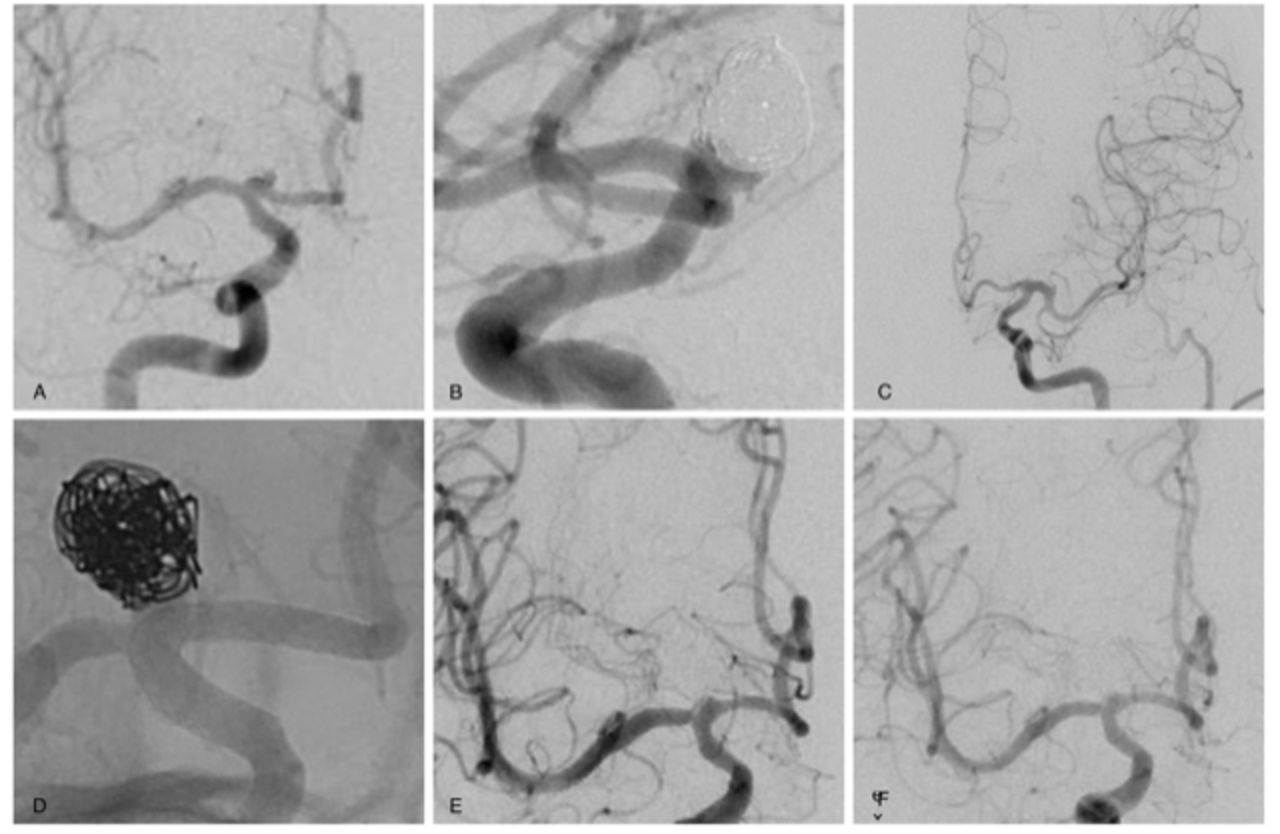
- Figure 1
Case 8: Cinquagenarian with (A,B) R ICA DSA showing growing residual of a previously coiled 8 mm right A1 aneurysm and (C) L CCA DSA showing absent ACoA. (D) Unsubtracted DSA during PED placement from R ACA into R ICA across M1 origin, which continued to fill anterograde, without delay, in parallel with the ACA at both (E) 6 months and (F) 12 months follow-up R ICA DSA. ACA, anterior cerebral artery; ACoA, anterior communicating artery; CCA, common carotid artery; DSA, digital subtraction angiography; ICA, internal carotid artery; PED, pipeline embolisation device.
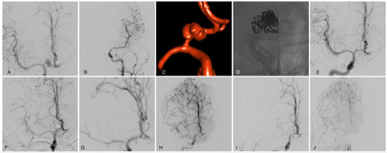
- Figure 2
Case 5: Sexagenarian with (A) R CCA DSA showing absent ACoA and (B) L CCA DSA showing fusiform PCoA aneurysm with (C) dome irregularity and short postcommunicating landing zone in the ICA. (D) Treatment with single-stage pipeline with adjunctive coiling from the ACA into the ICA. (E) Immediate postprocedural DSA showing platelet aggregation at the M1 origin and delayed MCA filling when the patient awoke with right upper extremity (RUE) weakness (F) resolved clinically and improved angiographically after intra-arterial Abciximab administration. Follow-up DSA at (G) 2 months, (H) 6 months, (I) 12 months, after which Prasugrel was weaned and (J) 24 months showing progressive recruitment of pial collaterals from the ACA and ECA to supply the MCA territory and moderately delayed filling of the covered MCA in the parenchymal phase. ACA, anterior cerebral artery; ACoA, anterior communicating artery; CCA, common carotid artery; DSA, digital subtraction angiography; ECA, external carotid artery; ICA, internal carotid artery; MCA, middle cerebral artery; PCoA, posterior communicating artery.
- Figure 3
Case 1: Teenage patient with history of bicoronal craniotomy for craniopharyngioma resection followed by proton beam therapy who presented with (A) R ICA DSA showing 11 mm A1 aneurysm and (B) L CCA DSA showing hypoplastic left ACA. (C) 3-D rotational angiography shows fusiform and highly irregular morphology. (D) Unsubtracted DSA from treatment with single-stage pipeline with adjunctive coiling from R ACA into R ICA. Follow-up R CCA DSA at (E) 2 months shows dome occlusion of the aneurysm with residual neck filling and anterograde arterial phase filling of the jailed R MCA. (F) 6 months shows some ghosting across the M1, (G,H) increased ghosting at 12 months follow-up DSA after stopping Plavix with significant recruitment of pial collaterals from the ACA in the late arterial and parenchymal phase. (I) 24-month DSA arterial phase shows limited anterograde filling of the jailed MCA and (J) robust pial collaterals from ACA and ECA apparent on parenchymal phase runs. ACA, anterior cerebral artery; CCA, common carotid artery; DSA, digital subtraction angiography; ECA, external carotid artery; ICA, internal carotid artery; MCA, middle cerebral artery.


Tables
- Table 1: Case details
Case no. Size (mm) Side Morphology Location Clinical history Treatment Complications Aneurysm occlusion MCA delay 1 11 R Fusiform A h/o craniopharyngioma rsxn, proton beam PED+coil – Complete Significant 2 6 R Fusiform PCoA, ICAT h/o SAH, prev coil/recur ICAT, new PCoA PED – Complete Minimal 3 9 L Saccular A1 Incidental PED+coil Retroperitoneal haematoma, L MCA stroke, mortality – –- 4 7 R Saccular A1 Incidental PED – Complete Minimal 5 17 L Fusiform PCoA h/o SAH from ruptured ACoA PED R paresis on emergence; DSA M1 platelet plug; resolved with Reopro Dome occlusion/
neck residualModerate 6 3 R Saccular A1 Incidental PED Post-embo hypotensions, MCA ischaemia, residual LUE weakness (mRS=2) Complete Significant 7 4 R Saccular A1 Incidental PED – Complete Moderate 8 8 R Saccular ICAT h/o coiling, recanalisation PED – Complete Minimal 9 10 R Pseudoaneurysm PCoA Traumatic ICA injury PED Intraprocedural platelet aggregation, resolved with Reopro Complete Minimal DSA, digital subtraction angiography; ICAT, internal carotid artery termination; MCA, middle cerebral artery; PCoA, posterior communicating artery; PED, pipeline embolisation device.


