

Article Figures & Data
Figures
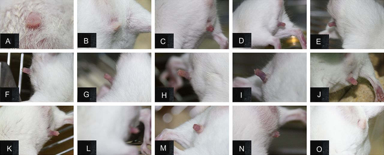
- Figure 1
Adult mice were found to display priapism after permanent middle cerebral artery occlusion (pMCAO). Photographs showed that adult mouse did not get erection before pMCAO (A), or 1 day after pMCAO (B). Persistent erection started on the 2nd day (C), maintained through the 3rd (D) to the 13th day (N) and disappeared on the 14th day postoperation (O).
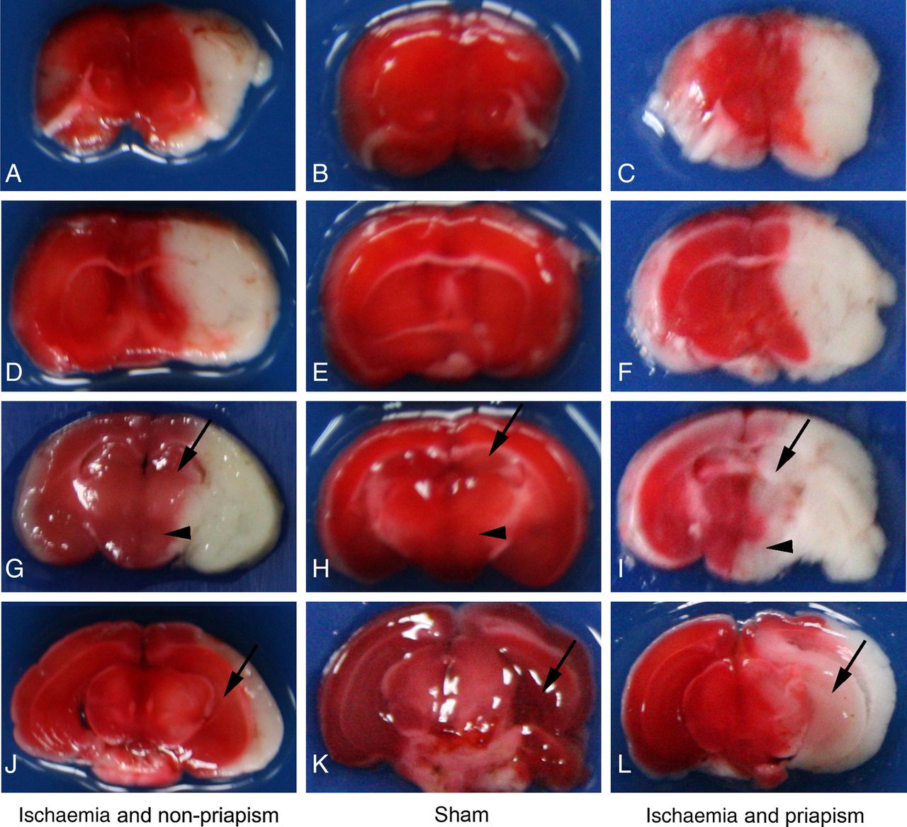
- Figure 2
Priapism closely correlated with infarct area. Photographs showed that the sham group did not get priapism and infarct in the brain (middle column, B–K); ischaemic mice that also displayed priapism were found to have extensive infarct affecting both hypothalamus (arrowhead) and hippocampus (arrow) areas (right column, C–L); in contrast, ischaemic mice that did not display priapism were found to have non-infarct hypothalamus and hippocampus areas (left column, A–J). All mice were sacrificed 3 days after permanent middle cerebral artery occlusion, n=4 for each group.
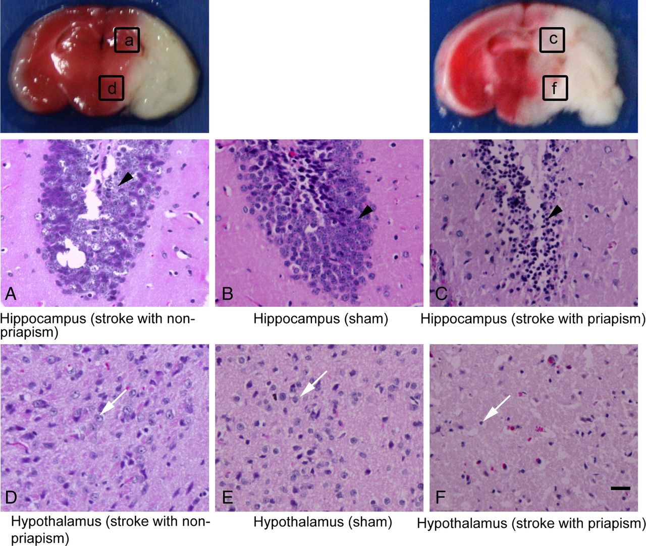
- Figure 3
Priapism occurrence was related with different infarct area. H&E staining showed that the sham group did not show priapism, and the nuclei of cells in these brains were normal (B and E); in the group of ischaemic mice with priapism (C and F), the nuclei of the cells in the hippocampus (arrow head) and hypothalamus (white arrow) displayed shrinkage; however, in the group of ischaemic mice without priapism (A and D), the hippocampus (arrowhead) and hypothalamus (white arrow) showed normal nuclei size; photographs A, C, D, F were taken from a, c, d and f, respectively. Bar=20 µm, n=4 for each group.
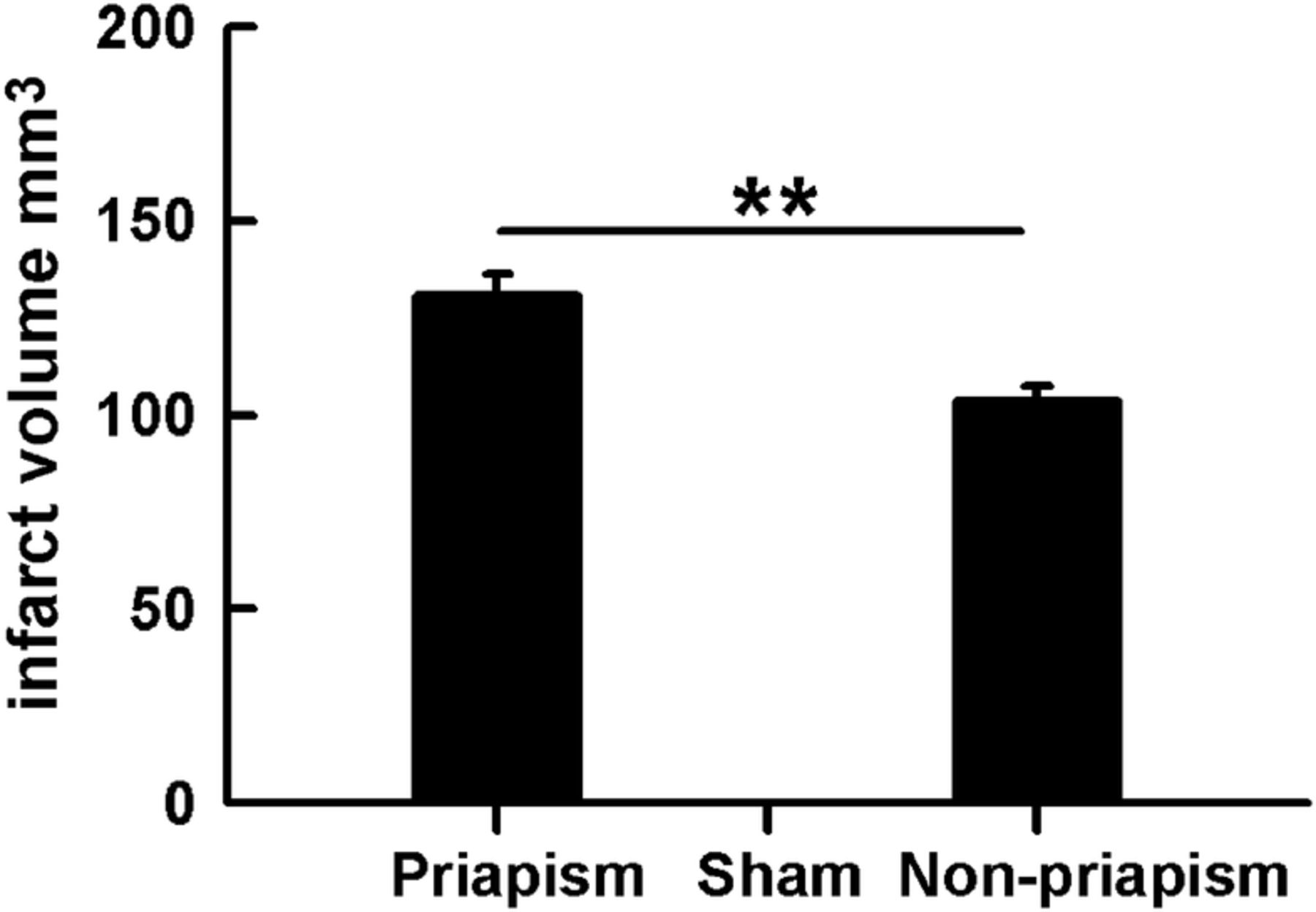
- Figure 4
The infarct volume was significantly larger in stroke mice with priapism than those without priapism. Bar graph showed significant difference of infarct volume between the stroke priapism mice and stroke non-priapism mice. **p<0.01, n=4 for each group.


Tables
- Table 1
A summary of priapism condition in nine mice after permanent middle cerebral artery occlusion (pMCAO)
Number Erection start (days) Erection disappear (days) 1 2 14 2 2 14 3 2 7 4 / / 5 / / 6 1 10 7 2 9 8 / / 9 1 11 The first line means nine mice were recorded after pMCAO (number 1–9), the middle and the last line displayed when priapism had occurred and disappeared after surgery. ‘/’ Indicates no priapism was observed.


