

Article Figures & Data
Figures
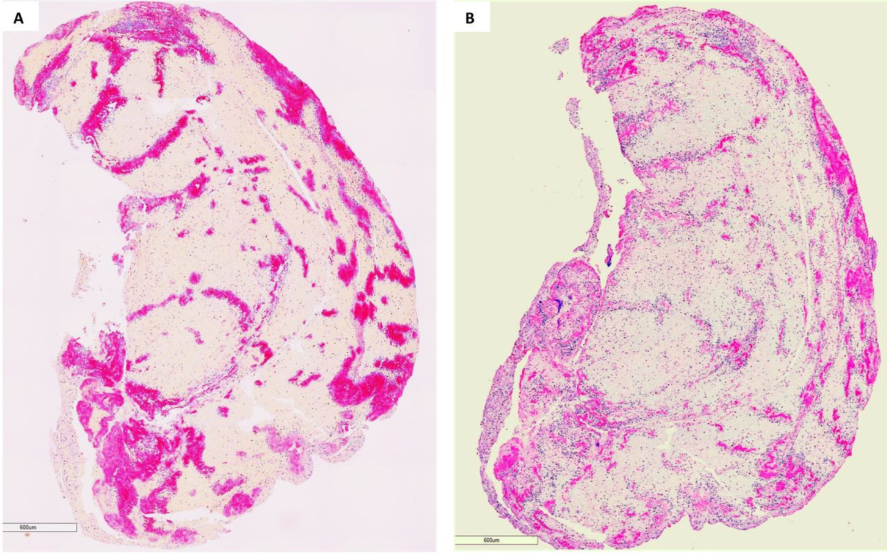
- Figure 1
Heterogeneous feature of stroke emboli. At least two serial sections of thrombi retrieved from ischaemic stroke patients were stained with Martius scarlet blue to identify different component of thrombus. (A–C) Representative microphotographs taken with lower magnification from three different patients’ thrombi, showing the thrombus tissue consists of different component which is stained with different colour (yellow, pinkish, red and blue). (D–F) Microphotographs taken with higher magnification from the rectangular area in (A–C), showing red blood cells (yellow), fibrin strands (red/ intense pink), platelets (pinkish) and white blood cells (blue) all are present.
- Figure 2
Colocalisation of platelets and Von Willebrand Factor (vWF). Serial sections of thrombi retrieved from ischaemic stroke patients were stained with antibodies against CD42b and vWF to visualise the copresence of platelets and vWF. (A and B) Representative macrophotographs of serial sections from one thrombus stained for CD42b (A) and vWF (B), showing the distribution (red/pink) of platelets and vWF are same (Immunohistochemistry, original magnification ×4.0).
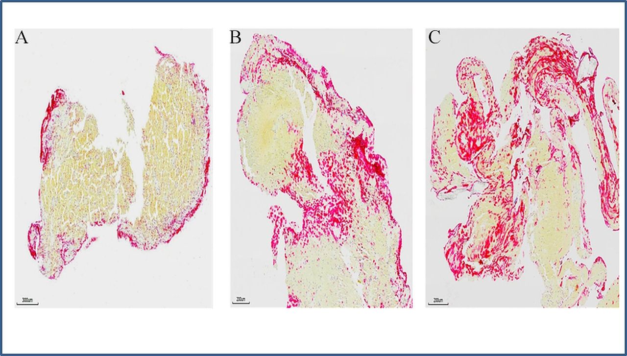
- Figure 3
Neutrophil extracellular traps (NETs) in stroke emboli. Serial sections of thrombi retrieved from ischaemic stroke patients were stained with antibody against citrullinated histone H3 (H3Cit) to reveal the presence of NETs. (A–C) Representative microphotographs of three different patients’ thrombi stained for H3Cit (red), showing NETs is expressed along the surface of thrombus (A), at junctions between RBC-rich and platelet rich-regions (B and C). (Immunohistochemistry, original magnification ×3.7 (A), ×4.9 (B and C)).
- Figure 4
Representative scanning electron microscopy (SEM) images of an acute ischaemic stroke thrombus. (A) Arrows correspond to the locations analysed by SEM. The surface of the clot shows multiple folds and ridges. (B) Distinct thrombotic components are not discernible on the surface that appears smooth. (C) High magnification of the surface suggestive of dense integration of components and advanced organisation. Only a few red blood cells are evident. (D) The rift on the surface shows the presence of a dense outer shell (arrow) and a different structure of the interior of the clot (asterisk). (E) Magnified area of the interior of the clot displays numerous individual polyhedrocytes and some distinct fibrin strands suggesting an immature structure. (F) The cross-section exposes the thrombus core which is detailed in (G). (H) Identifiable thrombus components indicate a limited maturity and incomplete integration. Scale bar=10 µm (C, E, H), 100 µm (B, D, G), 200 µm (F) and 500 µm (A).
- Figure 5
Three-dimensional scanning electron microscopy analysis in three representative cases. Volume renderings assembled from serial block-face imaging highlight detailed ultrastructural organisation and characteristics of individual components for each clot (red blood cells (RBC)=yellow arrows, fibrin=red arrows, white blood cells=blue arrow). (A) Clot area with tightly packed RBC as polyhedrocytes intermixed with a limited volume of thin fibrin fibres. (B) Clot area with mixed composition consisting of both packed polyhedrocytes and dense network of thick fibrin fibres. (C) Fibrin-rich area containing dense fibrin masses with sparse polyhedrocytes.


Tables
- Table 1
Summary table
Clot component Structure Interacts with Potential therapeutic agent(s) Fibrin (factor Ia) Fibrous protein Platelets, factor XIII, RBCs Alteplase, tenecteplase Platelets Nucleus-free cell fragments Collagen, vWF, WBCs GpIIb/IIIa blockers vWF Plasma glycoprotein Fibrin, platelets, collagen ADAMTS13, N-acetyle cysteine (NAC) NET Decondensed DNA/chromatin Thrombin, platelets, vWF DNase1 NET, neutrophil extracellular trap; RBC, red blood cell; vWF, von Willebrand Factor; WBC, white blood cell.


