
Main menu

  • Online first
    • Online first
  • Current issue
    • Current issue
  • Archive
    • Archive
  • Submit a paper
    • Online submission site
    • Instructions for authors
  • About the journal
    • About the journal
    • Editorial board
    • Instructions for authors
    • FAQs
    • Chinese Stroke Association
  • Help
    • Contact us
    • Feedback form
    • Reprints
    • Permissions
    • Advertising
  • BMJ Journals

User menu

  • Login

Search

  • Advanced search
  • BMJ Journals
  • Login
  • Facebook
  • Twitter
Stroke and Vascular Neurology

Advanced Search

  • Online first
    • Online first
  • Current issue
    • Current issue
  • Archive
    • Archive
  • Submit a paper
    • Online submission site
    • Instructions for authors
  • About the journal
    • About the journal
    • Editorial board
    • Instructions for authors
    • FAQs
    • Chinese Stroke Association
  • Help
    • Contact us
    • Feedback form
    • Reprints
    • Permissions
    • Advertising
Open Access

MRI and glymphatic system

Quan Jiang
DOI: 10.1136/svn-2018-000197 Published 4 July 2019
Quan Jiang
Departments of Neurology, Henry Ford Hospital, Detroit, Michigan, USA
  • Find this author on Google Scholar
  • Find this author on PubMed
  • Search for this author on this site
  • ORCID record for Quan Jiang
  • Article
  • Figures & Data
  • eLetters
  • Info & Metrics
  • PDF
Loading

Abstract

Glymphatic system is newly discovered pseudolymphatic system in brain and it plays an important role in the removal of interstitial metabolic waste products. We discuss and review the role of glymphatic system in neurological diseases, especially focus on the potential of MRI modelling and its associated challenges to obtain useful new information related to understand the glymphatic system dynamics, pathways, and provide quantitative maps for diagnosis, monitoring and prognosis of the disease. The non-invasive nature of MRI might more readily help with translation of glymphatic measurements from the laboratory to the clinic.

Introduction of glymphatic system

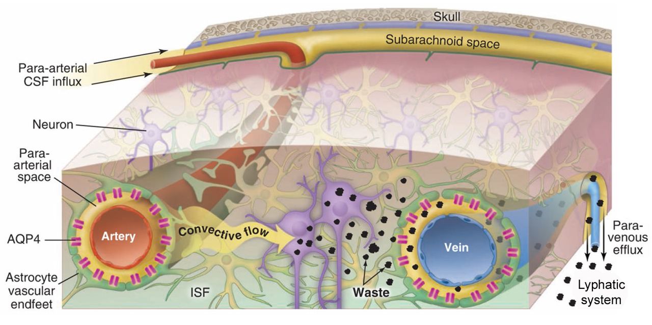
The traditional concept of cerebrospinal fluid (CSF) hydrodynamics is being challenged by recent publications.1–6 The traditional model of CSF believes that CSF is produced in the choroid plexus in four ventricles, then flows from the ventricles to the subarachnoid spaces, and from there flows out along spinal and cranial nerves, the arachnoid villi and the olfactory bulb. Recent investigations provide evidence showing that a newly identified system, the glymphatic system, exists in the brain which facilitates the exchange of interstitial fluid (ISF) and CSF, and provide a pathway of organised convective fluid flow to drive clearance of interstitial solutes with wastes from the brain.1–6 The glymphatic system consists of para-arterial CSF influx and paravenous efflux routes, which are dependent on the astrocytic aquaporin-4 (AQP4) water channel.1–5 As demonstrated in figure 1, the influx flow from subarachnoid CSF enters parenchymal through para-arterial pathways and then exchanges with ISF facilitated by AQP4, both are cleared together with associated solutes along paravenous pathways, from where it exits into head lymphatic vessels.3 Weller et al defined paravascular drainage pathways in the brain, and speculated about its influence on Aβ clearance from the brain.7 8 Iliff et al had demonstrated the greatest tracer accumulation was detected surrounding large-calibre draining veins.3 Louveau et al and Aspelund et al have recently discovered that the meninges of a lymphatic network drains the macromolecules and immune cells from the brain parenchyma.6 9

Figure 1
  • Download figure
  • Open in new tab
  • Download powerpoint
Figure 1

Influx, CSF-ISF exchange and efflux of glymphatic system: The influx flow of CSF into brain through the para-arterial pathways and then exchange with ISF. The mixed CSF and ISF with interstitial metabolic waste are flowed towards paravenous pathways, and then go to lymphatic system. AQP4 water channels in both arteries and veins play important role in reducing the resistance to CSF movement between paravascular spaces and the interstitium. Reproduced with permission from25. AQP4, astrocytic aquaporin-4; CSF, cerebrospinal fluid; ISF, interstitial fluid.

Glymphatic system and neurological diseases

The glymphatic system plays an important role in neurological diseases.1–5 10 Xie et al demonstrated that natural sleep or anaesthesia significantly enlarged the interstitial space which increased convective exchange of CSF with ISF and clearance rate of β-amyloid during sleep.1 Ageing strongly relates to many neurodegenerative diseases and is also associated with a slow waste clearance in the brain.11 CSF tracer clearance was gradually reduced from middle-aged (12–13 months) to old (18 months) brain compared with young (2–3 months) brain.11 In mouse Alzheimer’s disease model, Peng et al found reduced clearance of β-amyloid and glymphatic influx which worsened with advanced age.10 Reduced waste clearance has also been demonstrated after traumatic brain injury, haemorrhage, hypertension, acute ischaemia and multiple microinfarction.5 12–16 In diabetes mellitus (DM) animal model, tracer clearance from brain parenchyma was significantly reduced with diabetes, leading to accumulation of tracers in multiple brain regions which correlated with degree of cognitive decline.17 Although glymphatic system has exhibited its important role in neurological diseases, further investigation is necessary to develop glymphatic directed therapeutic strategies.

MRI detection and modelling of glymphatic system

Most previous studies of the glymphatic system have been performed with two-photon laser scanning microscopy, which is precise and excellent for a predetermined small paravascular space but not a suitable tool for whole brain study, especially deep brain tissues and also invasive. MRI overcomes the weak points of two-photon confocal microscopy and provides non-invasive whole brain dynamic real-time imaging of the glymphatic system. Iliff et al first employed MRI to monitor glymphatic function and demonstrated that MRI allowed the identification of glymphatic pathways, several influx nodes and some kinetic parameters to evaluate solute clearance in the brain.18 MRI has also exhibited its advantage to monitor whole brain pictures of slow waste clearance after subarachnoid haemorrhage and ischaemic stroke.13 15 16 A recent study has demonstrated that compared with middle-aged non-DM animals, tracer clearance from brain parenchyma measured by both MRI and fluorescence imaging was significantly reduced with DM.17 MRI cluster analysis measurement of glymphatic system also exhibits a significant increase in paravascular space in the DM animals compared with control animals. Most interestingly, the MRI measures of tracer clearance, residual intensity and paravascular space are highly sensitive indices of pathological changes occurring early during the development of DM compared with functional behavioural tests and vascular dysfunction.

Although MRI measurements of glymphatic system have exhibited a great potential for non-invasive evaluation of whole brain glymphatic functions, properly modelling this newly discovered system is still challenge. So far, few studies have been published for modelling glymphatic system. Ratner et al 19 20 identified the glymphatic pathways by using optimal mass transport to model the glymphatic flow vector field. Results from this study agreed with previous results of the glymphatic pathways. However, a weak point of this model is no quantitative measures of this system. Lee et al performed a very interesting study using kinetic modelling to quantify CSF-ISF exchange rates in supine, prone or lateral positions. This model exhibited the ability to differential the CSF-ISF exchange rates in three different postures and the quantitative maps derived from this model showed that glymphatic transport was most efficient in the lateral position compared with the supine or prone positions.21 One limitation for this model is its global input function which could produce more errors, especially for glymphatic system.

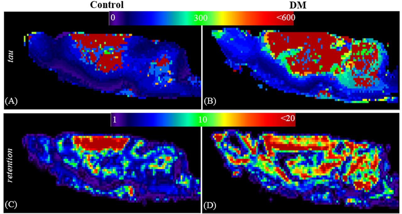
A newly published model using local input function has exhibited its advantage for improving the detecting sensitivity of glymphatic system.22 This model employed cluster analysis to determine the similar regions with the time signal curves from dynamic contrast agent MRI data and then derive local input function based on signal exchanges between regions.22 The results from this new model exhibit its better sensitivity to differential diabetic and control animals than previous two-compartment kinetic model and show increased clearance time and binding of tracers in DM compared with control animal brain (figure 2). Arterial input using global input function is a long-standing issue and the major source of error in cerebral permeability and perfusion measurements. Although model-based approach had tried to improve the delay and dispersion issues and to derive the local input function in permeability and perfusion measurements,23 24 directly measure local input function is still a challenge. Compared with vascular system, the errors caused global input delay and dispersion in glymphatic system are more severe due to much longer delay and tissue involved dispersion. The newly published model22 provides a better approach for more accurate evaluate glymphatic changes under neurological diseases.

Figure 2
  • Download figure
  • Open in new tab
  • Download powerpoint
Figure 2

The maps of quantitative modelling the glymphatic system for control and DM animals. Figure 2 shows the visual comparison of the clearance time constant, tau (minutes, (A, B) and the retention (C, D) maps under control (A, C) and DM (B, D) conditions. Comparing with the control, DM animal exhibits longer clearance time constant (slower clearance of the tracer from the tissue) and high retention values (large fraction of the tracer remains in brain tissue). DM, diabetes mellitus.

Current MRI modelling of glymphatic system only considers a pressure-driven motion of bulk fluid referred to as ‘advection’. The waste clearance could also involve diffusion although it will play much less role in the waste clearance. The further modelling of brain waste clearance could include both advection and diffusion to further reduce system errors.

Conclusion

Glymphatic system plays important role in neurological diseases. MRI could provide more information about glymphatic functions in whole brain study, especially deep brain tissues. Precisely modelling glymphatic system could obtain useful new information related to understand the glymphatic system dynamics, pathways, and provide quantitative maps for diagnosis, monitoring and prognosis of the disease.

Acknowledgments

This work was supported by NIH grants RF1 AG057494, RO1 NS108463, RO1 NS097747 and R21 AG052735. I thank Li Zhang, Esmaeil Davoodi-Bojd, Guangliang Ding, Zhenggang Zhang and Michael Chopp for their contribution of the glymphatic studies in the Department of Neurology, Henry Ford Hospital.

Footnotes

  • Contributors QJ is the sole author of the contribution.

  • Funding The authors have not declared a specific grant for this research from any funding agency in the public, commercial or not-for-profit sectors.

  • Competing interests None declared.

  • Provenance and peer review Commissioned; externally peer reviewed.

  • Patient consent for publication Not required.

This is an open access article distributed in accordance with the Creative Commons Attribution Non Commercial (CC BY-NC 4.0) license, which permits others to distribute, remix, adapt, build upon this work non-commercially, and license their derivative works on different terms, provided the original work is properly cited, appropriate credit is given, any changes made indicated, and the use is non-commercial. See: http://creativecommons.org/licenses/by-nc/4.0/.

References

  1. 1.↵
    1. Xie L ,
    2. Kang H ,
    3. Xu Q , et al
    . Sleep drives metabolite clearance from the adult brain. Science 2013;342:373–7.doi:10.1126/science.1241224
    OpenUrlAbstract/FREE Full Text
  2. 2.↵
    1. Iliff JJ ,
    2. Wang M ,
    3. Zeppenfeld DM , et al
    . Cerebral arterial pulsation drives paravascular CSF-interstitial fluid exchange in the murine brain. J Neurosci 2013;33:18190–9.doi:10.1523/JNEUROSCI.1592-13.2013
    OpenUrlAbstract/FREE Full Text
  3. 3.↵
    1. Iliff JJ ,
    2. Wang M ,
    3. Liao Y , et al
    . A paravascular pathway facilitates CSF flow through the brain parenchyma and the clearance of interstitial solutes, including amyloid β. Sci Transl Med 20124:ra111.doi:10.1126/scitranslmed.3003748
    OpenUrl
  4. 4.↵
    1. Rangroo Thrane V ,
    2. Thrane AS ,
    3. Plog BA , et al
    . Paravascular microcirculation facilitates rapid lipid transport and astrocyte signaling in the brain. Sci Rep 2013;3:2582.doi:10.1038/srep02582
    OpenUrlCrossRefPubMed
  5. 5.↵
    1. Plog BA ,
    2. Dashnaw ML ,
    3. Hitomi E , et al
    . Biomarkers of traumatic injury are transported from brain to blood via the glymphatic system. J Neurosci 2015;35:518–26.doi:10.1523/JNEUROSCI.3742-14.2015
    OpenUrlAbstract/FREE Full Text
  6. 6.↵
    1. Louveau A ,
    2. Smirnov I ,
    3. Keyes TJ , et al
    . Structural and functional features of central nervous system lymphatic vessels. Nature 2015;523:337–41.doi:10.1038/nature14432
    OpenUrlCrossRefPubMed
  7. 7.↵
    1. Pollock H ,
    2. Hutchings M ,
    3. Weller RO , et al
    . Perivascular spaces in the basal ganglia of the human brain: their relationship to lacunes. J Anat 1997;191:337–46.doi:10.1046/j.1469-7580.1997.19130337.x
    OpenUrlCrossRefPubMed
  8. 8.↵
    1. Weller RO ,
    2. Kida S ,
    3. Zhang ET
    . Pathways of fluid drainage from the brain-morphological aspects and immunological significance in rat and man. Brain Pathol 1992;2:277–84.doi:10.1111/j.1750-3639.1992.tb00704.x
    OpenUrlCrossRefPubMedWeb of Science
  9. 9.↵
    1. Aspelund A ,
    2. Antila S ,
    3. Proulx ST , et al
    . A dural lymphatic vascular system that drains brain interstitial fluid and macromolecules. J Exp Med 2015;212:991–9.doi:10.1084/jem.20142290
    OpenUrlAbstract/FREE Full Text
  10. 10.↵
    1. Peng W ,
    2. Achariyar TM ,
    3. Li B , et al
    . Suppression of glymphatic fluid transport in a mouse model of Alzheimer’s disease. Neurobiol Dis 2016;93:215–25.doi:10.1016/j.nbd.2016.05.015
    OpenUrl
  11. 11.↵
    1. Kress BT ,
    2. Iliff JJ ,
    3. Xia M , et al
    . Impairment of paravascular clearance pathways in the aging brain. Ann Neurol 2014;76:845–61.doi:10.1002/ana.24271
    OpenUrlCrossRefPubMed
  12. 12.↵
    1. Iliff JJ ,
    2. Chen MJ ,
    3. Plog BA , et al
    . Impairment of glymphatic pathway function promotes tau pathology after traumatic brain injury. J Neurosci 2014;34:16180–93.doi:10.1523/JNEUROSCI.3020-14.2014
    OpenUrlAbstract/FREE Full Text
  13. 13.↵
    1. Wang M ,
    2. Ding F ,
    3. Deng S , et al
    . Focal solute trapping and global glymphatic pathway impairment in a murine model of multiple microinfarcts. J Neurosci 2017;37:2870–7.doi:10.1523/JNEUROSCI.2112-16.2017
    OpenUrlAbstract/FREE Full Text
  14. 14.↵
    1. Mestre H ,
    2. Tithof J ,
    3. Du T , et al
    . Flow of cerebrospinal fluid is driven by arterial pulsations and is reduced in hypertension. Nat Commun 2018;9:4878.doi:10.1038/s41467-018-07318-3
    OpenUrlCrossRef
  15. 15.↵
    1. Goulay R ,
    2. Flament J ,
    3. Gauberti M , et al
    . Subarachnoid hemorrhage severely impairs brain parenchymal cerebrospinal fluid circulation in nonhuman primate. Stroke 2017;48:2301–5.doi:10.1161/STROKEAHA.117.017014
    OpenUrlAbstract/FREE Full Text
  16. 16.↵
    1. Gaberel T ,
    2. Gakuba C ,
    3. Goulay R , et al
    . Impaired glymphatic perfusion after strokes revealed by contrast-enhanced MRI: a new target for fibrinolysis? Stroke 2014;45:3092–6.doi:10.1161/STROKEAHA.114.006617
    OpenUrlAbstract/FREE Full Text
  17. 17.↵
    1. Jiang Q ,
    2. Zhang L ,
    3. Ding G , et al
    . Impairment of the glymphatic system after diabetes. J Cereb Blood Flow Metab 2017;37:1326–37.doi:10.1177/0271678X16654702
    OpenUrl
  18. 18.↵
    1. Iliff JJ ,
    2. Lee H ,
    3. Yu M , et al
    . Brain-wide pathway for waste clearance captured by contrast-enhanced MRI. J Clin Invest 2013;123:1299–309.doi:10.1172/JCI67677
    OpenUrlCrossRefPubMedWeb of Science
  19. 19.↵
    1. Ratner V ,
    2. Gao Y ,
    3. Lee H , et al
    . Cerebrospinal and interstitial fluid transport via the glymphatic pathway modeled by optimal mass transport. Neuroimage 2017;152:530–7.doi:10.1016/j.neuroimage.2017.03.021
    OpenUrl
  20. 20.↵
    1. Ratner V ,
    2. Zhu L ,
    3. Kolesov I , et al
    . Optimal-mass-transfer-based estimation of glymphatic transport in living brain. Proc SPIE Int Soc Opt Eng 2015;9413:9413.doi:10.1117/12.2076289
    OpenUrl
  21. 21.↵
    1. Lee H ,
    2. Xie L ,
    3. Yu M , et al
    . The effect of body posture on brain glymphatic transport. J Neurosci 2015;35:11034–44.doi:10.1523/JNEUROSCI.1625-15.2015
    OpenUrlAbstract/FREE Full Text
  22. 22.↵
    1. Davoodi-Bojd E ,
    2. Ding G ,
    3. Zhang L , et al
    . Modeling glymphatic system of the brain using MRI. Neuroimage 2018;188:616–27.doi:10.1016/j.neuroimage.2018.12.039
    OpenUrl
  23. 23.↵
    1. Nejad-Davarani SP ,
    2. Bagher-Ebadian H ,
    3. Ewing JR , et al
    . An extended vascular model for less biased estimation of permeability parameters in DCE-T1 images. NMR Biomed 2017;30:e3698.doi:10.1002/nbm.3698
    OpenUrl
  24. 24.↵
    1. Nejad-Davarani SP ,
    2. Bagher-Ebadian H ,
    3. Ewing JR , et al
    . A parametric model of the brain vascular system for estimation of the arterial input function (AIF) at the tissue level. NMR Biomed 2017;30:e3695.doi:10.1002/nbm.3695
    OpenUrl
  25. 25.↵
    1. Nedergaard M
    . Garbage Truck of the Brain. Science 2013;340:1529–30.doi:10.1126/science.1240514
    OpenUrlAbstract/FREE Full Text
PreviousNext
Back to top
Vol 4 Issue 2 Table of Contents
Stroke and Vascular Neurology: 4 (2)
  • Table of Contents
  • About the Cover
  • Index by author
Email

Thank you for your interest in spreading the word on Stroke and Vascular Neurology.

NOTE: We only request your email address so that the person you are recommending the page to knows that you wanted them to see it, and that it is not junk mail. We do not capture any email address.

Enter multiple addresses on separate lines or separate them with commas.
MRI and glymphatic system
(Your Name) has sent you a message from Stroke and Vascular Neurology
(Your Name) thought you would like to see the Stroke and Vascular Neurology web site.
CAPTCHA
This question is for testing whether or not you are a human visitor and to prevent automated spam submissions.
Print
Alerts
Sign In to Email Alerts with your Email Address
Citation Tools
MRI and glymphatic system
Quan Jiang
Stroke and Vascular Neurology Jun 2019, 4 (2) 75-77; DOI: 10.1136/svn-2018-000197

Citation Manager Formats

  • BibTeX
  • Bookends
  • EasyBib
  • EndNote (tagged)
  • EndNote 8 (xml)
  • Medlars
  • Mendeley
  • Papers
  • RefWorks Tagged
  • Ref Manager
  • RIS
  • Zotero
Cite This
  • APA
  • Chicago
  • Endnote
  • MLA
Loading
MRI and glymphatic system
Quan Jiang
Stroke and Vascular Neurology Jun 2019, 4 (2) 75-77; DOI: 10.1136/svn-2018-000197
Download PDF

Share
MRI and glymphatic system
Quan Jiang
Stroke and Vascular Neurology Jun 2019, 4 (2) 75-77; DOI: 10.1136/svn-2018-000197
Reddit logo Twitter logo Facebook logo Mendeley logo
Respond to this article
  • Tweet Widget
  • Facebook Like
  • Google Plus One
  • Article
    • Abstract
    • Introduction of glymphatic system
    • Glymphatic system and neurological diseases
    • MRI detection and modelling of glymphatic system
    • Conclusion
    • Acknowledgments
    • Footnotes
    • References
  • Figures & Data
  • eLetters
  • Info & Metrics
  • PDF

Related Articles

Cited By...

More in this TOC Section

  • Design of trials in lacunar stroke and cerebral small vessel disease: review and experience with the LACunar Intervention Trial 2 (LACI-2)
  • Anti-stroke biologics: from recombinant proteins to stem cells and organoids
  • Central post-stroke pain: advances in clinical and preclinical research
Show more Review

Similar Articles


CONTENT

  • Latest content
  • Current issue
  • Archive
  • eLetters
  • Sign up for email alerts
  • RSS

JOURNAL

  • About the journal
  • Editorial board
  • Recommend to librarian
  • Chinese Stroke Association

AUTHORS

  • Instructions for authors
  • Submit a paper
  • Track your article
  • Open Access at BMJ

HELP

  • Contact us
  • Reprints
  • Permissions
  • Advertising
  • Feedback form

© 2025 Chinese Stroke Association