
Main menu

  • Online first
    • Online first
  • Current issue
    • Current issue
  • Archive
    • Archive
  • Submit a paper
    • Online submission site
    • Instructions for authors
  • About the journal
    • About the journal
    • Editorial board
    • Instructions for authors
    • FAQs
    • Chinese Stroke Association
  • Help
    • Contact us
    • Feedback form
    • Reprints
    • Permissions
    • Advertising
  • BMJ Journals

User menu

  • Login

Search

  • Advanced search
  • BMJ Journals
  • Login
  • Facebook
  • Twitter
Stroke and Vascular Neurology

Advanced Search

  • Online first
    • Online first
  • Current issue
    • Current issue
  • Archive
    • Archive
  • Submit a paper
    • Online submission site
    • Instructions for authors
  • About the journal
    • About the journal
    • Editorial board
    • Instructions for authors
    • FAQs
    • Chinese Stroke Association
  • Help
    • Contact us
    • Feedback form
    • Reprints
    • Permissions
    • Advertising
Open Access

Cerebral microvascular dysfunction and neurodegeneration in dementia

Feng Han
DOI: 10.1136/svn-2018-000213 Published 4 July 2019
Feng Han
Key Laboratory of Cardiovascular and Cerebrovascular Medicine, School of Pharmacy, Nanjing Medical University, Nanjing, Jiangsu, China
  • Find this author on Google Scholar
  • Find this author on PubMed
  • Search for this author on this site
  • ORCID record for Feng Han
  • Article
  • Figures & Data
  • eLetters
  • Info & Metrics
  • PDF
Loading

Abstract

Maintaining normal learning and memory functions requires a high degree of coordination between neural and vascular cells. Basic and clinical studies have shown that brain microvasculature dysfunction activates inflammatory cells in the brain, leading to progressive neuronal loss and eventually dementia. This review focuses on recent studies aimed at identifying the molecular events that link cerebral microvascular dysfunction to neurodegeneration, including oxidative/nitrosative stress, cellular metabolic dysfunction, inflammatory signalling and abnormal synaptic plasticity. A better understanding of the coupling between vasculature and brain neurons and how this coupling is disrupted under pathological conditions is of great significance in identifying new diagnostic and treatment targets for dementia for which no new drugs have been approved since 2003.

Accumulating evidence from epidemiological and postmortem studies suggests a close relationship among small vessel disease (SVD), aberrant neurovascular regulation and cognitive impairment. Dysregulation of cerebral blood flow and abnormal neural activity secondary to altered neurovascular coupling have been observed in the early stage of neurodegenerative diseases. However, the precise mechanism by which vascular dysfunction contributes to cognitive impairment remains poorly understood.

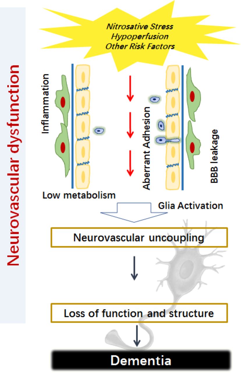
Aberrant peroxynitrite generation is a primary risk factor for vascular dysfunction.1–4 Patients with Alzheimer’s disease (AD) exhibit clear cerebrovascular pathology and white matter injury due to microvascular infarction, suggesting an association of the cerebrovascular disease with neurodegenerative disorders.5 6 A possible contribution of nitrosative stress to AD pathology is indicated by the observation that peroxynitrite formation induced microvascular injury followed by β-amyloid accumulation in microvessels and parenchyma and eventual Glycogen synthase kinase-3β (GSK3β) activation and hyperphosphorylation of tau proteins in the aged rat brain.5 Supporting this suggestion, inhibition of nitrosative stress partially restored the decrease of phospho-CaMKII (Th286/287) and phospho-synapsin I (Ser603), increased the number of mature neurons in the hippocampus, and significantly improved cognitive function in mice with vascular dementia.7 Moreover, the inhibition markedly decreased peroxynitrite formation and downregulated NLRP3/caspase-1/IL-1β signalling in mice with bilateral carotid artery stenosis (figure 1).8

Figure 1
  • Download figure
  • Open in new tab
  • Download powerpoint
Figure 1

The schematic image is illustrating the possible relationship between cerebral microvascular dysfunction and neurodegeneration in dementia.

Damages to the neurovascular system are believed to be a major source of morbidity and mortality in chronic diseases such as diabetes.9 An alternation in hippocampal Ca2+/calmodulin-dependent protein kinase II(CaMKII)/Protein kinase C(PKC)/Protein kinase A(PKA) pathway may be partially responsible for the detrimental postdiabetic outcomes associated with cognitive dysfunction in diabetic models.10 Brain microvascular complications can lead to changes in brain structure and function.11 12 In vivo two-photon fluorescence microscopy reveals disturbances of cerebral capillary blood flow in diabetes which are exaggerated and associated with rapid cognitive decline after brain ischaemia.11 Accompanied by the disturbance of capillary blood flow and neurovascular damage, phospho-CaMKII (Thr286), phospho-synapsin I (Ser603) and phospho-GluR1 (Ser831) are dramatically decreased in the diabetic mice with ischaemia.11 Together, these findings suggest an active role for vascular factors in the pathogenesis of cognitive dysfunction. Further studies are needed to fully understand the changes in the microenvironment in the brain under the different pathological condition and the communication between components of the neurovascular unit.

Emerging studies have uncovered essential roles of endothelial molecules in brain function and behaviour.13–17 For example, brain endothelial cells have been suggested to be a natural gatekeeper for virus-induced sickness behaviour.18 The study further demonstrated an engagement of tissue-specific Interferon (IFN) receptor chain 1 and established the signal transduction axis as a target for the treatment of the behavioural changes.18 Clinical studies suggest that endothelial thrombomodulin is closely associated with cerebral SVD severity, but cerebral endothelial activation in deep penetrating arteries did not.19 By using both endothelial-specific gene knockdown and neurobehavioural strategies, we found that deletion of the endothelial ErbB4 receptor-induced impairment in exploratory activity in adult mice and the effect was associated with reduced glucose transport and impaired energy metabolism of the brain.20 More recently, the study showed that brain endothelial-derived semaphorin 3G might act as a synaptic organiser, regulating synaptic plasticity and hippocampal-dependent memory.21

A long list of endeavours are warranted to further tackle the mechanisms underlying the interaction between vascular and neuronal cells and the role of the interaction in dementia, including (1) studies using novel technologies including imaging from nanoscale to whole brain, high-throughput transcriptomics/proteomics screens and bioinformatics, (2) a deeper understanding of the dynamic regulation of neurovascular coupling by circulating factors including those implicated in dementia, and (3) further exploration of the role of exosomes, cytokines and microRNAs in neurovascular compartments. A combination of these approaches may allow for multidimensional analyses of the complex underlying molecular and cellular network, help discover novel biomarkers, and develop more effective diagnostic and treatment strategies for dementia.

Footnotes

  • Contributors FH is the sole author.

  • Funding This work was supported by the State Key Program of National Natural Science of China (grant number 81730101), the National Key Research and Development Program of China (grant number 2016YFE0125400), and the National Natural Science Foundation of China (grant number 81573411).

  • Competing interests None declared.

  • Provenance and peer review Commissioned; externally peer reviewed.

  • Data sharing statement No additional data are available.

  • Patient consent for publication Not required.

This is an open access article distributed in accordance with the Creative Commons Attribution Non Commercial (CC BY-NC 4.0) license, which permits others to distribute, remix, adapt, build upon this work non-commercially, and license their derivative works on different terms, provided the original work is properly cited, appropriate credit is given, any changes made indicated, and the use is non-commercial. See: http://creativecommons.org/licenses/by-nc/4.0/.

References

  1. 1.↵
    1. Han F ,
    2. Tao RR ,
    3. Zhang GS , et al
    . Melatonin ameliorates ischemic-like injury-evoked nitrosative stress: Involvement of HtrA2/PED pathways in endothelial cells. J Pineal Res 2011;50:281–91.doi:10.1111/j.1600-079X.2010.00838.x
    OpenUrlPubMed
  2. 2.↵
    1. Tao RR ,
    2. Ji YL ,
    3. Lu YM , et al
    . Targeting nitrosative stress for neurovascular protection: new implications in brain diseases. Curr Drug Targets 2012;13:272–84.doi:10.2174/138945012799201649
    OpenUrlCrossRefPubMedWeb of Science
  3. 3.↵
    1. Han F ,
    2. Chen YX ,
    3. Lu YM ,
    4. Ym L , et al
    . Regulation of the ischemia-induced autophagy-lysosome processes by nitrosative stress in endothelial cells. J Pineal Res 2011;51:124–35.doi:10.1111/j.1600-079X.2011.00869.x
    OpenUrlCrossRefPubMed
  4. 4.↵
    1. Wang CK ,
    2. Ahmed MM ,
    3. Jiang Q , et al
    . Melatonin ameliorates hypoglycemic stress-induced brain endothelial tight junction injury by inhibiting protein nitration of TP53-induced glycolysis and apoptosis regulator. J Pineal Res 2017;63:e12440.doi:10.1111/jpi.12440
    OpenUrl
  5. 5.↵
    1. Han F ,
    2. Ali Raie A ,
    3. Shioda N , et al
    . Accumulation of beta-amyloid in the brain microvessels accompanies increased hyperphosphorylated tau proteins following microsphere embolism in aged rats. Neuroscience 2008;153:414–27.doi:10.1016/j.neuroscience.2008.02.044
    OpenUrl
  6. 6.↵
    1. Miklossy J
    . Cerebral hypoperfusion induces cortical watershed microinfarcts which may further aggravate cognitive decline in Alzheimer’s disease. Neurol Res 2003;25:605–10.doi:10.1179/016164103101202048
    OpenUrlCrossRefPubMed
  7. 7.↵
    1. Wang XJ ,
    2. Gao YP ,
    3. Lu NN , et al
    . Endogenous Polysialic Acid Based Micelles for Calmodulin Antagonist Delivery against Vascular Dementia. ACS Appl Mater Interfaces 2016;8:35045–58.doi:10.1021/acsami.6b13052
    OpenUrl
  8. 8.↵
    1. Wang R ,
    2. Yin YX ,
    3. Mahmood Q , et al
    . Calmodulin inhibitor ameliorates cognitive dysfunction via inhibiting nitrosative stress and NLRP3 signaling in mice with bilateral carotid artery stenosis. CNS Neurosci Ther 2017;23:818–26.doi:10.1111/cns.12726
    OpenUrl
  9. 9.↵
    1. Bogush M ,
    2. Heldt NA ,
    3. Persidsky Y
    . Blood Brain Barrier Injury in Diabetes: Unrecognized Effects on Brain and Cognition. J Neuroimmune Pharmacol 2017;12:593–601.doi:10.1007/s11481-017-9752-7
    OpenUrl
  10. 10.↵
    1. Liao MH ,
    2. Xiang YC ,
    3. Huang JY , et al
    . The disturbance of hippocampal CaMKII/PKA/PKC phosphorylation in early experimental diabetes mellitus. CNS Neurosci Ther 2013;19:329–36.doi:10.1111/cns.12084
    OpenUrl
  11. 11.↵
    1. Huang JY ,
    2. Li LT ,
    3. Wang H , et al
    . In vivo two-photon fluorescence microscopy reveals disturbed cerebral capillary blood flow and increased susceptibility to ischemic insults in diabetic mice. CNS Neurosci Ther 2014;20:816–22.doi:10.1111/cns.12268
    OpenUrlCrossRefPubMed
  12. 12.↵
    1. Mogi M ,
    2. Horiuchi M
    . Neurovascular coupling in cognitive impairment associated with diabetes mellitus. Circ J 2011;75:1042–8.doi:10.1253/circj.CJ-11-0121
    OpenUrlCrossRefPubMedWeb of Science
  13. 13.↵
    1. Toth P ,
    2. Tarantini S ,
    3. Csiszar A , et al
    . Functional vascular contributions to cognitive impairment and dementia: mechanisms and consequences of cerebral autoregulatory dysfunction, endothelial impairment, and neurovascular uncoupling in aging. Am J Physiol Heart Circ Physiol 2017;312:H1–H20.doi:10.1152/ajpheart.00581.2016
    OpenUrlCrossRefPubMed
  14. 14.↵
    1. Obasi CN ,
    2. Cruickshanks KJ ,
    3. Nondahl DM , et al
    . Association of Biomarkers for Inflammation, Endothelial Dysfunction and Oxidative Stress with Cognitive Impairment. The Epidemiology of Hearing Loss Study (EHLS). Oxid Antioxid Med Sci 2012;1:169–73.doi:10.5455/oams.031212.br.004
    OpenUrlPubMed
  15. 15.↵
    1. Kinzenbaw DA ,
    2. Chu Y ,
    3. Peña Silva RA , et al
    . Interleukin-10 protects against aging-induced endothelial dysfunction. Physiol Rep 2013;1:e00149.doi:10.1002/phy2.149
    OpenUrlAbstract/FREE Full Text
  16. 16.↵
    1. Cahill-Smith S ,
    2. Li JM
    . Oxidative stress, redox signalling and endothelial dysfunction in ageing-related neurodegenerative diseases: a role of NADPH oxidase 2. Br J Clin Pharmacol 2014;78:441–53.doi:10.1111/bcp.12357
    OpenUrlCrossRefPubMed
  17. 17.↵
    1. Fujiyoshi K ,
    2. Yamaoka-Tojo M ,
    3. Minami Y , et al
    . Endothelial Dysfunction Is Associated with Cognitive Impairment of Elderly Cardiovascular Disease Patients. Int Heart J 2018;59:1034–40.doi:10.1536/ihj.17-610
    OpenUrl
  18. 18.↵
    1. Blank T ,
    2. Detje CN ,
    3. Spieß A , et al
    . Brain Endothelial- and Epithelial-Specific Interferon Receptor Chain 1 Drives Virus-Induced Sickness Behavior and Cognitive Impairment. Immunity 2016;44:901–12.doi:10.1016/j.immuni.2016.04.005
    OpenUrl
  19. 19.↵
    1. Giwa MO ,
    2. Williams J ,
    3. Elderfield K , et al
    . Neuropathologic evidence of endothelial changes in cerebral small vessel disease. Neurology 2012;78:167–74.doi:10.1212/WNL.0b013e3182407968
    OpenUrlCrossRefPubMed
  20. 20.↵
    1. Wu G ,
    2. Liu XX ,
    3. Lu NN ,
    4. Nn L , et al
    . Endothelial ErbB4 deficit induces alterations in exploratory behavior and brain energy metabolism in mice. CNS Neurosci Ther 2017;23:510–7.doi:10.1111/cns.12695
    OpenUrl
  21. 21.↵
    1. Tan C ,
    2. Lu NN ,
    3. Wang CK , et al
    . Endothelium-Derived Semaphorin 3G Regulates Hippocampal Synaptic Structure and Plasticity via Neuropilin-2/PlexinA4. Neuron 2019;101:920–37.doi:10.1016/j.neuron.2018.12.036
    OpenUrl
PreviousNext
Back to top
Vol 4 Issue 2 Table of Contents
Stroke and Vascular Neurology: 4 (2)
  • Table of Contents
  • About the Cover
  • Index by author
Email

Thank you for your interest in spreading the word on Stroke and Vascular Neurology.

NOTE: We only request your email address so that the person you are recommending the page to knows that you wanted them to see it, and that it is not junk mail. We do not capture any email address.

Enter multiple addresses on separate lines or separate them with commas.
Cerebral microvascular dysfunction and neurodegeneration in dementia
(Your Name) has sent you a message from Stroke and Vascular Neurology
(Your Name) thought you would like to see the Stroke and Vascular Neurology web site.
CAPTCHA
This question is for testing whether or not you are a human visitor and to prevent automated spam submissions.
Print
Alerts
Sign In to Email Alerts with your Email Address
Citation Tools
Cerebral microvascular dysfunction and neurodegeneration in dementia
Feng Han
Stroke and Vascular Neurology Jun 2019, 4 (2) 105-107; DOI: 10.1136/svn-2018-000213

Citation Manager Formats

  • BibTeX
  • Bookends
  • EasyBib
  • EndNote (tagged)
  • EndNote 8 (xml)
  • Medlars
  • Mendeley
  • Papers
  • RefWorks Tagged
  • Ref Manager
  • RIS
  • Zotero
Cite This
  • APA
  • Chicago
  • Endnote
  • MLA
Loading
Cerebral microvascular dysfunction and neurodegeneration in dementia
Feng Han
Stroke and Vascular Neurology Jun 2019, 4 (2) 105-107; DOI: 10.1136/svn-2018-000213
Download PDF

Share
Cerebral microvascular dysfunction and neurodegeneration in dementia
Feng Han
Stroke and Vascular Neurology Jun 2019, 4 (2) 105-107; DOI: 10.1136/svn-2018-000213
Reddit logo Twitter logo Facebook logo Mendeley logo
Respond to this article
  • Tweet Widget
  • Facebook Like
  • Google Plus One
  • Article
    • Abstract
    • Footnotes
    • References
  • Figures & Data
  • eLetters
  • Info & Metrics
  • PDF

Related Articles

Cited By...

More in this TOC Section

  • Design of trials in lacunar stroke and cerebral small vessel disease: review and experience with the LACunar Intervention Trial 2 (LACI-2)
  • Anti-stroke biologics: from recombinant proteins to stem cells and organoids
  • Central post-stroke pain: advances in clinical and preclinical research
Show more Review

Similar Articles


CONTENT

  • Latest content
  • Current issue
  • Archive
  • eLetters
  • Sign up for email alerts
  • RSS

JOURNAL

  • About the journal
  • Editorial board
  • Recommend to librarian
  • Chinese Stroke Association

AUTHORS

  • Instructions for authors
  • Submit a paper
  • Track your article
  • Open Access at BMJ

HELP

  • Contact us
  • Reprints
  • Permissions
  • Advertising
  • Feedback form

© 2025 Chinese Stroke Association