
Main menu

  • Online first
    • Online first
  • Current issue
    • Current issue
  • Archive
    • Archive
  • Submit a paper
    • Online submission site
    • Instructions for authors
  • About the journal
    • About the journal
    • Editorial board
    • Instructions for authors
    • FAQs
    • Chinese Stroke Association
  • Help
    • Contact us
    • Feedback form
    • Reprints
    • Permissions
    • Advertising
  • BMJ Journals

User menu

  • Login

Search

  • Advanced search
  • BMJ Journals
  • Login
  • Facebook
  • Twitter
Stroke and Vascular Neurology

Advanced Search

  • Online first
    • Online first
  • Current issue
    • Current issue
  • Archive
    • Archive
  • Submit a paper
    • Online submission site
    • Instructions for authors
  • About the journal
    • About the journal
    • Editorial board
    • Instructions for authors
    • FAQs
    • Chinese Stroke Association
  • Help
    • Contact us
    • Feedback form
    • Reprints
    • Permissions
    • Advertising
Open Access

Update on cerebral small vessel disease: a dynamic whole-brain disease

Yulu Shi, Joanna M Wardlaw
DOI: 10.1136/svn-2016-000035 Published 25 October 2016
Yulu Shi
1Centre for Clinical Brain Sciences, University of Edinburgh, Edinburgh, UK
2Department of Neurology, Zhongnan Hospital, Wuhan University, Wuhan, China
  • Find this author on Google Scholar
  • Find this author on PubMed
  • Search for this author on this site
Joanna M Wardlaw
1Centre for Clinical Brain Sciences, University of Edinburgh, Edinburgh, UK
  • Find this author on Google Scholar
  • Find this author on PubMed
  • Search for this author on this site
  • Article
  • Figures & Data
  • eLetters
  • Info & Metrics
  • PDF
Loading

Article Figures & Data

Figures

  • Additional Files
  • Figure 1
    • Download figure
    • Open in new tab
    • Download powerpoint
    Figure 1

    STRIVE, STandards for Reporting and Imaging of Small Vessel Disease: example findings (upper), schematic representation (middle) and a summary of imaging characteristics (lower) of MRI features for changes related to small vessel disease.4 DWI, diffusion-weighted imaging; FLAIR, fluid-attenuated inversion recovery; SWI, susceptibility-weighted imaging; GRE, gradient-recalled echo.
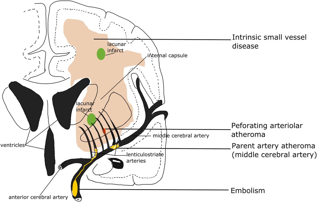
  • Figure 2
    • Download figure
    • Open in new tab
    • Download powerpoint
    Figure 2

    Four possible mechanisms that cause a lacunar infarct (from bottom to top): (A) an embolus from the big arteries or cardiac sources goes up to MCA and ends up entering and occluding lenticulostriate arteries, resulting in a lacunar lesion in basal ganglia; (B) if the atheroma in the parent artery (ie, MCA) is positioned at the opening of its penetrating branches, it could lead to an acute occlusion of one or several penetrating arteries, hence causing a lacunar infarct; (C) a lacunar infarct could also be due to atheroma in the perforating artery if an acute occlusion happens; (D) intrinsic small vessel disease may lead to diffused disrupted blood–brain barrier. If this happens at an arteriolar level, plasma fluid components would enter and deposit in the vessel wall, resulting in narrowing of the arteriolar lumen, vessel wall thickening and eventually a secondary luminal occlusion and traditional infarct. MCA, middle cerebral arteries.
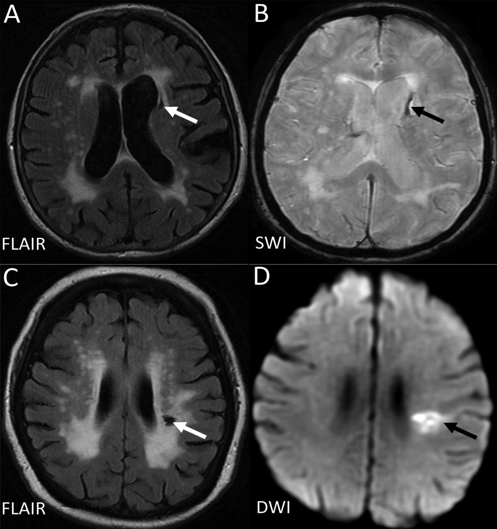
  • Figure 3
    • Download figure
    • Open in new tab
    • Download powerpoint
    Figure 3

    Example of MRIs of a lacune from a haemorrhagic source (A,B), and from a lacunar infarct (C, D). D (the DWI) is from the acute presentation (i.e. within a few days of the stroke), and C (the FlAIR) is weeks to months later when the lesion has cavitated. DWI, diffusion-weighted imaging; FLAIR, fluid-attenuated inversion recovery; SWI, susceptibility-weighted imaging.
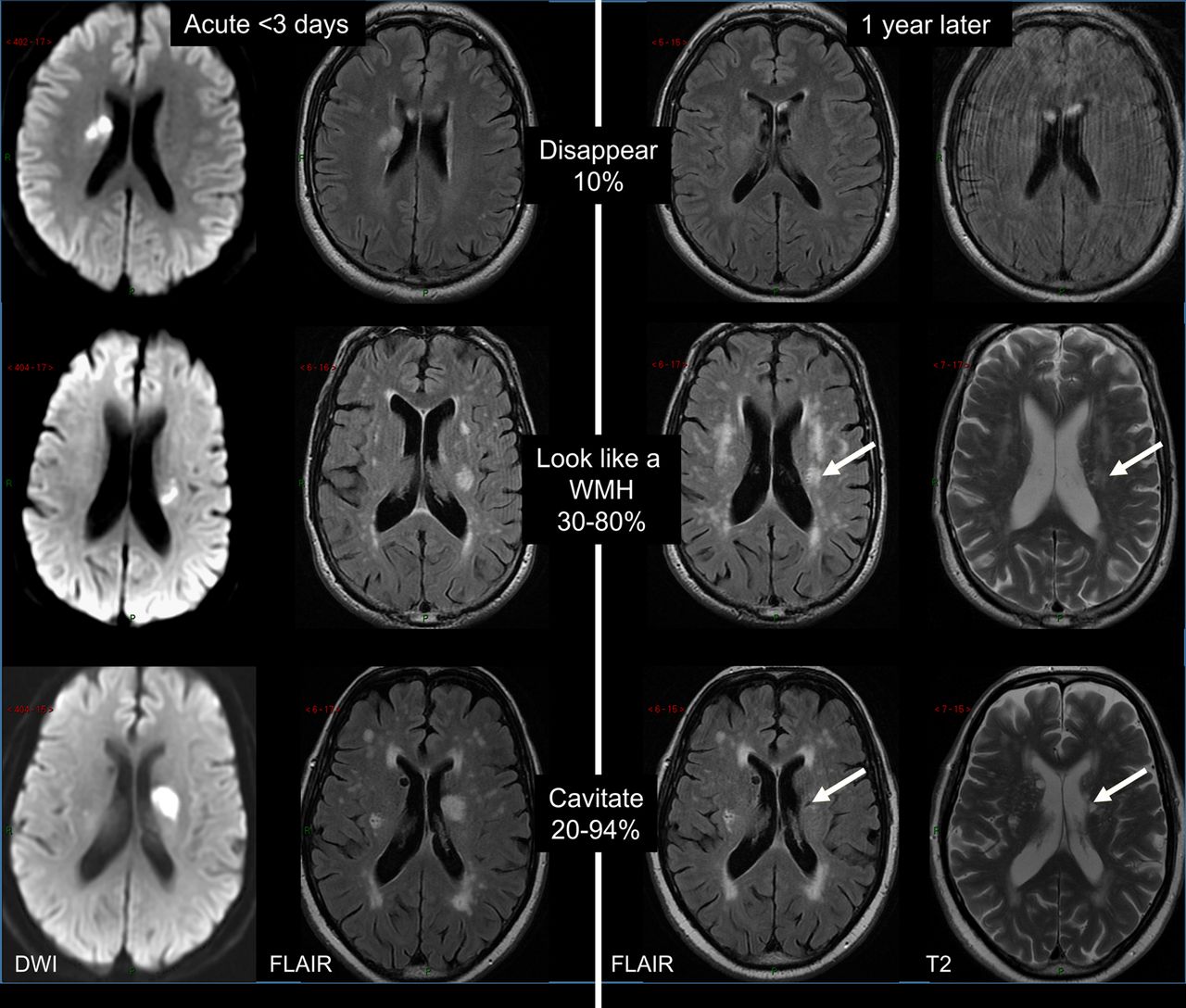
  • Figure 4
    • Download figure
    • Open in new tab
    • Download powerpoint
    Figure 4

    Long-term appearances of lacunar infarcts (arrows: old stroke lesion on the follow-up scans). DWI, diffusion-weighted imaging; FLAIR, fluid-attenuated inversion recovery; WMH, white matter hyperintensity.

Additional Files

  • Figures
  • Abstract in Chinese

    This web only file has been produced by the BMJ Publishing Group from an electronic file supplied by the author(s) and has not been edited for content.

    • Abstract in Chinese - Online abstract
PreviousNext
Back to top
Vol 1 Issue 3 Table of Contents
Stroke and Vascular Neurology: 1 (3)
  • Table of Contents
  • About the Cover
  • Index by author
Email

Thank you for your interest in spreading the word on Stroke and Vascular Neurology.

NOTE: We only request your email address so that the person you are recommending the page to knows that you wanted them to see it, and that it is not junk mail. We do not capture any email address.

Enter multiple addresses on separate lines or separate them with commas.
Update on cerebral small vessel disease: a dynamic whole-brain disease
(Your Name) has sent you a message from Stroke and Vascular Neurology
(Your Name) thought you would like to see the Stroke and Vascular Neurology web site.
CAPTCHA
This question is for testing whether or not you are a human visitor and to prevent automated spam submissions.
Print
Alerts
Sign In to Email Alerts with your Email Address
Citation Tools
Update on cerebral small vessel disease: a dynamic whole-brain disease
Yulu Shi, Joanna M Wardlaw
Stroke and Vascular Neurology Sep 2016, 1 (3) 83-92; DOI: 10.1136/svn-2016-000035

Citation Manager Formats

  • BibTeX
  • Bookends
  • EasyBib
  • EndNote (tagged)
  • EndNote 8 (xml)
  • Medlars
  • Mendeley
  • Papers
  • RefWorks Tagged
  • Ref Manager
  • RIS
  • Zotero
Cite This
  • APA
  • Chicago
  • Endnote
  • MLA
Loading
Update on cerebral small vessel disease: a dynamic whole-brain disease
Yulu Shi, Joanna M Wardlaw
Stroke and Vascular Neurology Sep 2016, 1 (3) 83-92; DOI: 10.1136/svn-2016-000035
Download PDF

Share
Update on cerebral small vessel disease: a dynamic whole-brain disease
Yulu Shi, Joanna M Wardlaw
Stroke and Vascular Neurology Sep 2016, 1 (3) 83-92; DOI: 10.1136/svn-2016-000035
Reddit logo Twitter logo Facebook logo Mendeley logo
Respond to this article
  • Tweet Widget
  • Facebook Like
  • Google Plus One
  • Article
    • Abstract
    • Introduction
    • Footnotes
    • References
  • Figures & Data
  • eLetters
  • Info & Metrics
  • PDF

Related Articles

Cited By...

More in this TOC Section

  • Design of trials in lacunar stroke and cerebral small vessel disease: review and experience with the LACunar Intervention Trial 2 (LACI-2)
  • Anti-stroke biologics: from recombinant proteins to stem cells and organoids
  • Central post-stroke pain: advances in clinical and preclinical research
Show more Review

Similar Articles


CONTENT

  • Latest content
  • Current issue
  • Archive
  • eLetters
  • Sign up for email alerts
  • RSS

JOURNAL

  • About the journal
  • Editorial board
  • Recommend to librarian
  • Chinese Stroke Association

AUTHORS

  • Instructions for authors
  • Submit a paper
  • Track your article
  • Open Access at BMJ

HELP

  • Contact us
  • Reprints
  • Permissions
  • Advertising
  • Feedback form

© 2025 Chinese Stroke Association